

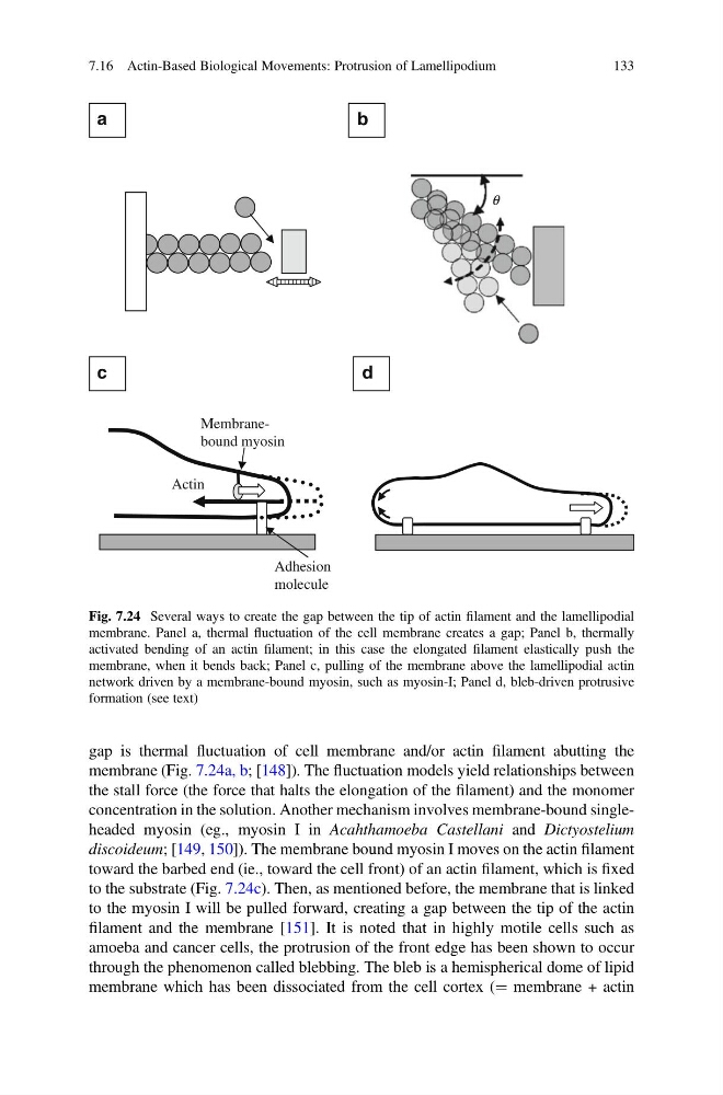
gap is thermal fluctuation of cell membrane and/or actin filament abutting the
membrane (Fig. 7.24a, b; [148]). The fluctuation models yield relationships between
the stall force (the force that halts the elongation of the filament) and the monomer
concentration in the solution. Another mechanism involves membrane-bound singleheaded myosin (eg., myosin I in Acahthamoeba Castellani and Dictyostelium
discoideum; [149, 150]). The membrane bound myosin I moves on the actin filament
toward the barbed end (ie., toward the cell front) of an actin filament, which is fixed
to the substrate (Fig. 7.24c). Then, as mentioned before, the membrane that is linked
to the myosin I will be pulled forward, creating a gap between the tip of the actin
filament and the membrane [151]. It is noted that in highly motile cells such as
amoeba and cancer cells, the protrusion of the front edge has been shown to occur
through the phenomenon called blebbing. The bleb is a hemispherical dome of lipid
membrane which has been dissociated from the cell cortex (¼ membrane + actin
a
c
d
b
Actin
Membranebound myosin
Adhesion
molecule
q
Fig. 7.24 Several ways to create the gap between the tip of actin filament and the lamellipodial
membrane. Panel a, thermal fluctuation of the cell membrane creates a gap; Panel b, thermally
activated bending of an actin filament; in this case the elongated filament elastically push the
membrane, when it bends back; Panel c, pulling of the membrane above the lamellipodial actin
network driven by a membrane-bound myosin, such as myosin-I; Panel d, bleb-driven protrusive
formation (see text)
7.16 Actin-Based Biological Movements: Protrusion of Lamellipodium
133
membrane (Fig. 7.24a, b; [148]). The fluctuation models yield relationships between
the stall force (the force that halts the elongation of the filament) and the monomer
concentration in the solution. Another mechanism involves membrane-bound singleheaded myosin (eg., myosin I in Acahthamoeba Castellani and Dictyostelium
discoideum; [149, 150]). The membrane bound myosin I moves on the actin filament
toward the barbed end (ie., toward the cell front) of an actin filament, which is fixed
to the substrate (Fig. 7.24c). Then, as mentioned before, the membrane that is linked
to the myosin I will be pulled forward, creating a gap between the tip of the actin
filament and the membrane [151]. It is noted that in highly motile cells such as
amoeba and cancer cells, the protrusion of the front edge has been shown to occur
through the phenomenon called blebbing. The bleb is a hemispherical dome of lipid
membrane which has been dissociated from the cell cortex (¼ membrane + actin
a
c
d
b
Actin
Membranebound myosin
Adhesion
molecule
q
Fig. 7.24 Several ways to create the gap between the tip of actin filament and the lamellipodial
membrane. Panel a, thermal fluctuation of the cell membrane creates a gap; Panel b, thermally
activated bending of an actin filament; in this case the elongated filament elastically push the
membrane, when it bends back; Panel c, pulling of the membrane above the lamellipodial actin
network driven by a membrane-bound myosin, such as myosin-I; Panel d, bleb-driven protrusive
formation (see text)
7.16 Actin-Based Biological Movements: Protrusion of Lamellipodium
133