

rhodamine, [39] or prodan [40]; upon polymerization, those fluorophores are thought
to be buried in the interface between adjacent protomers and quantum yield increases
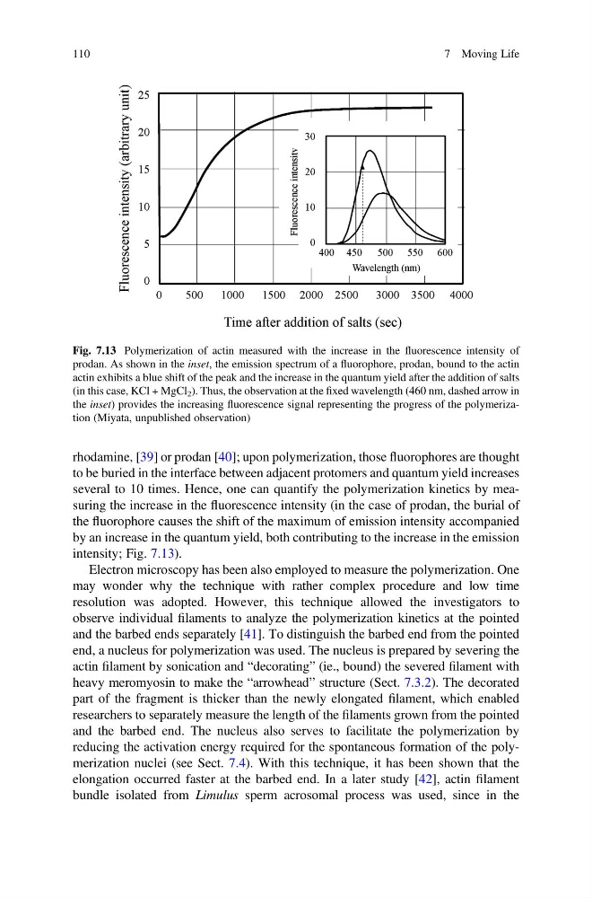
several to 10 times. Hence, one can quantify the polymerization kinetics by measuring the increase in the fluorescence intensity (in the case of prodan, the burial of
the fluorophore causes the shift of the maximum of emission intensity accompanied
by an increase in the quantum yield, both contributing to the increase in the emission
intensity; Fig. 7.13).
Electron microscopy has been also employed to measure the polymerization. One
may wonder why the technique with rather complex procedure and low time
resolution was adopted. However, this technique allowed the investigators to
observe individual filaments to analyze the polymerization kinetics at the pointed
and the barbed ends separately [41]. To distinguish the barbed end from the pointed
end, a nucleus for polymerization was used. The nucleus is prepared by severing the
actin filament by sonication and “decorating” (ie., bound) the severed filament with
heavy meromyosin to make the “arrowhead” structure (Sect. 7.3.2). The decorated
part of the fragment is thicker than the newly elongated filament, which enabled
researchers to separately measure the length of the filaments grown from the pointed
and the barbed end. The nucleus also serves to facilitate the polymerization by
reducing the activation energy required for the spontaneous formation of the polymerization nuclei (see Sect. 7.4). With this technique, it has been shown that the
elongation occurred faster at the barbed end. In a later study [42], actin filament
bundle isolated from Limulus sperm acrosomal process was used, since in the
Fig. 7.13 Polymerization of actin measured with the increase in the fluorescence intensity of
prodan. As shown in the inset, the emission spectrum of a fluorophore, prodan, bound to the actin
actin exhibits a blue shift of the peak and the increase in the quantum yield after the addition of salts
(in this case, KCl + MgCl 2 ). Thus, the observation at the fixed wavelength (460 nm, dashed arrow in
the inset) provides the increasing fluorescence signal representing the progress of the polymerization (Miyata, unpublished observation)
110
7 Moving Life
to be buried in the interface between adjacent protomers and quantum yield increases
several to 10 times. Hence, one can quantify the polymerization kinetics by measuring the increase in the fluorescence intensity (in the case of prodan, the burial of
the fluorophore causes the shift of the maximum of emission intensity accompanied
by an increase in the quantum yield, both contributing to the increase in the emission
intensity; Fig. 7.13).
Electron microscopy has been also employed to measure the polymerization. One
may wonder why the technique with rather complex procedure and low time
resolution was adopted. However, this technique allowed the investigators to
observe individual filaments to analyze the polymerization kinetics at the pointed
and the barbed ends separately [41]. To distinguish the barbed end from the pointed
end, a nucleus for polymerization was used. The nucleus is prepared by severing the
actin filament by sonication and “decorating” (ie., bound) the severed filament with
heavy meromyosin to make the “arrowhead” structure (Sect. 7.3.2). The decorated
part of the fragment is thicker than the newly elongated filament, which enabled
researchers to separately measure the length of the filaments grown from the pointed
and the barbed end. The nucleus also serves to facilitate the polymerization by
reducing the activation energy required for the spontaneous formation of the polymerization nuclei (see Sect. 7.4). With this technique, it has been shown that the
elongation occurred faster at the barbed end. In a later study [42], actin filament
bundle isolated from Limulus sperm acrosomal process was used, since in the
Fig. 7.13 Polymerization of actin measured with the increase in the fluorescence intensity of
prodan. As shown in the inset, the emission spectrum of a fluorophore, prodan, bound to the actin
actin exhibits a blue shift of the peak and the increase in the quantum yield after the addition of salts
(in this case, KCl + MgCl 2 ). Thus, the observation at the fixed wavelength (460 nm, dashed arrow in
the inset) provides the increasing fluorescence signal representing the progress of the polymerization (Miyata, unpublished observation)
110
7 Moving Life