

7.3.3 Actin Polymerization and Depolymerization
7.3.3.1 Methods to Measure Polymerization/Depolymerization
Dynamics of polymerization and depolymerization and its regulation by a number of
actin binding proteins are highly important aspects in the dynamical changes of the
cell. Therefore, quantitative in vitro measurement of the polymerization/depolymerization kinetics of actin has been developed. In the early stage of the study, the
degree of polymerization has been measured by several methods such as flowinduced birefringence of actin filament solution [34] and light scattering at
90 degrees [35, 36]. The drawback of these methods is that they are not sensitive
to detect the growth of filament in the initial stage of polymerization, where the
length of the filament is < ~0.1 μm [35]. Liberation of Pi can be also used as an
indicator of the progress of polymerization [37], but the time resolution (at most a
few seconds) is low. Thus, one needs methodology with high sensitivity to the
growth of the filament and time resolution for the estimation of the kinetic parameters. The most common methodology is based on fluorometry, which is highly
sensitive to the filament growth with relatively high time resolution. In these
methods, actin monomers are labeled with fluorophores such as pyrene [38],
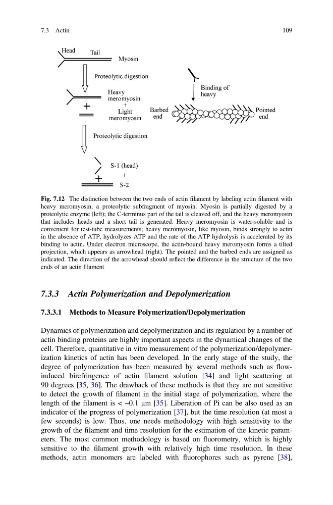
Fig. 7.12 The distinction between the two ends of actin filament by labeling actin filament with
heavy meromyosin, a proteolytic subfragment of myosin. Myosin is partially digested by a
proteolytic enzyme (left); the C-terminus part of the tail is cleaved off, and the heavy meromyosin
that includes heads and a short tail is generated. Heavy meromyosin is water-soluble and is
convenient for test-tube measurements; heavy meromyosin, like myosin, binds strongly to actin
in the absence of ATP, hydrolyzes ATP and the rate of the ATP hydrolysis is accelerated by its
binding to actin. Under electron microscope, the actin-bound heavy meromyosin forms a tilted
projection, which appears as arrowhead (right). The pointed and the barbed ends are assigned as
indicated. The direction of the arrowhead should reflect the difference in the structure of the two
ends of an actin filament
7.3 Actin
109
7.3.3.1 Methods to Measure Polymerization/Depolymerization
Dynamics of polymerization and depolymerization and its regulation by a number of
actin binding proteins are highly important aspects in the dynamical changes of the
cell. Therefore, quantitative in vitro measurement of the polymerization/depolymerization kinetics of actin has been developed. In the early stage of the study, the
degree of polymerization has been measured by several methods such as flowinduced birefringence of actin filament solution [34] and light scattering at
90 degrees [35, 36]. The drawback of these methods is that they are not sensitive
to detect the growth of filament in the initial stage of polymerization, where the
length of the filament is < ~0.1 μm [35]. Liberation of Pi can be also used as an
indicator of the progress of polymerization [37], but the time resolution (at most a
few seconds) is low. Thus, one needs methodology with high sensitivity to the
growth of the filament and time resolution for the estimation of the kinetic parameters. The most common methodology is based on fluorometry, which is highly
sensitive to the filament growth with relatively high time resolution. In these
methods, actin monomers are labeled with fluorophores such as pyrene [38],
Fig. 7.12 The distinction between the two ends of actin filament by labeling actin filament with
heavy meromyosin, a proteolytic subfragment of myosin. Myosin is partially digested by a
proteolytic enzyme (left); the C-terminus part of the tail is cleaved off, and the heavy meromyosin
that includes heads and a short tail is generated. Heavy meromyosin is water-soluble and is
convenient for test-tube measurements; heavy meromyosin, like myosin, binds strongly to actin
in the absence of ATP, hydrolyzes ATP and the rate of the ATP hydrolysis is accelerated by its
binding to actin. Under electron microscope, the actin-bound heavy meromyosin forms a tilted
projection, which appears as arrowhead (right). The pointed and the barbed ends are assigned as
indicated. The direction of the arrowhead should reflect the difference in the structure of the two
ends of an actin filament
7.3 Actin
109