

7.1.3 Single-Molecule Measurement for the Motor Function
of Myosin
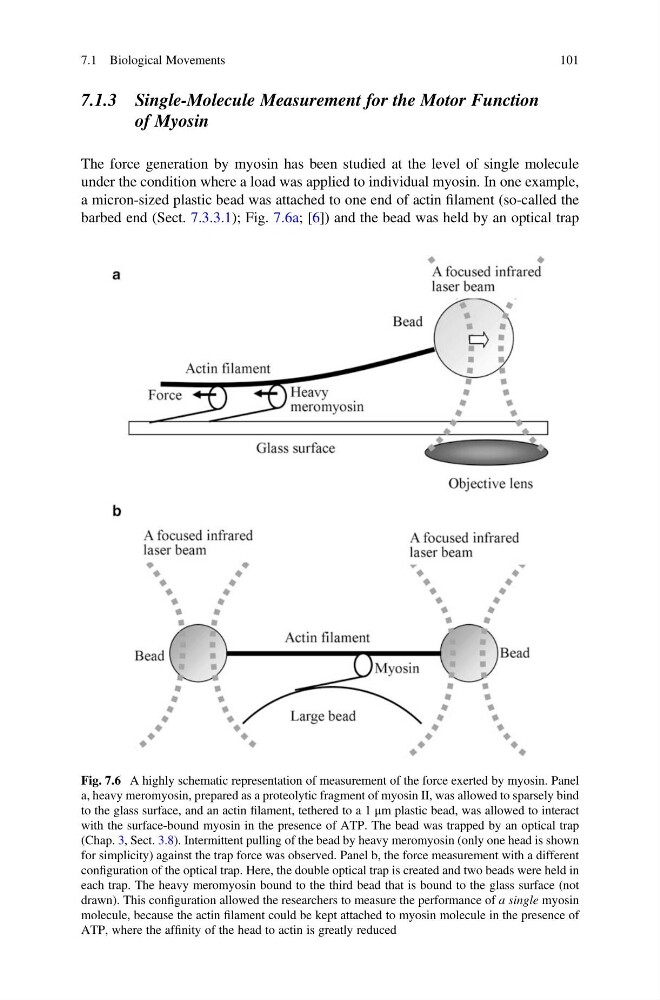
The force generation by myosin has been studied at the level of single molecule
under the condition where a load was applied to individual myosin. In one example,
a micron-sized plastic bead was attached to one end of actin filament (so-called the
barbed end (Sect. 7.3.3.1); Fig. 7.6a; [6]) and the bead was held by an optical trap
Fig. 7.6 A highly schematic representation of measurement of the force exerted by myosin. Panel
a, heavy meromyosin, prepared as a proteolytic fragment of myosin II, was allowed to sparsely bind
to the glass surface, and an actin filament, tethered to a 1 μm plastic bead, was allowed to interact
with the surface-bound myosin in the presence of ATP. The bead was trapped by an optical trap
(Chap. 3, Sect. 3.8). Intermittent pulling of the bead by heavy meromyosin (only one head is shown
for simplicity) against the trap force was observed. Panel b, the force measurement with a different
configuration of the optical trap. Here, the double optical trap is created and two beads were held in
each trap. The heavy meromyosin bound to the third bead that is bound to the glass surface (not
drawn). This configuration allowed the researchers to measure the performance of a single myosin
molecule, because the actin filament could be kept attached to myosin molecule in the presence of
ATP, where the affinity of the head to actin is greatly reduced
7.1 Biological Movements
101
of Myosin
The force generation by myosin has been studied at the level of single molecule
under the condition where a load was applied to individual myosin. In one example,
a micron-sized plastic bead was attached to one end of actin filament (so-called the
barbed end (Sect. 7.3.3.1); Fig. 7.6a; [6]) and the bead was held by an optical trap
Fig. 7.6 A highly schematic representation of measurement of the force exerted by myosin. Panel
a, heavy meromyosin, prepared as a proteolytic fragment of myosin II, was allowed to sparsely bind
to the glass surface, and an actin filament, tethered to a 1 μm plastic bead, was allowed to interact
with the surface-bound myosin in the presence of ATP. The bead was trapped by an optical trap
(Chap. 3, Sect. 3.8). Intermittent pulling of the bead by heavy meromyosin (only one head is shown
for simplicity) against the trap force was observed. Panel b, the force measurement with a different
configuration of the optical trap. Here, the double optical trap is created and two beads were held in
each trap. The heavy meromyosin bound to the third bead that is bound to the glass surface (not
drawn). This configuration allowed the researchers to measure the performance of a single myosin
molecule, because the actin filament could be kept attached to myosin molecule in the presence of
ATP, where the affinity of the head to actin is greatly reduced
7.1 Biological Movements
101