

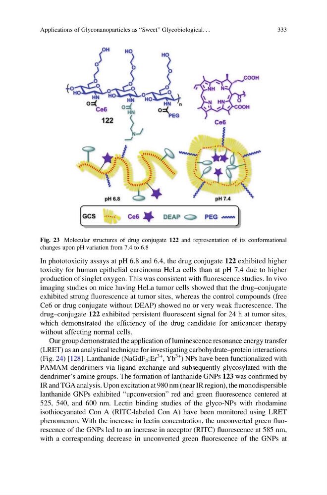
In phototoxicity assays at pH 6.8 and 6.4, the drug conjugate 122 exhibited higher
toxicity for human epithelial carcinoma HeLa cells than at pH 7.4 due to higher
production of singlet oxygen. This was consistent with fluorescence studies. In vivo
imaging studies on mice having HeLa tumor cells showed that the drug–conjugate
exhibited strong fluorescence at tumor sites, whereas the control compounds (free
Ce6 or drug conjugate without DEAP) showed no or very weak fluorescence. The
drug–conjugate 122 exhibited persistent fluorescent signal for 24 h at tumor sites,
which demonstrated the efficiency of the drug candidate for anticancer therapy
without affecting normal cells.
Our group demonstrated the application of luminescence resonance energy transfer
(LRET) as an analytical technique for investigating carbohydrate–protein interactions
(Fig. 24) [128]. Lanthanide (NaGdF 4 :Er
3+
, Yb
3+
) NPs have been functionalized with
PAMAM dendrimers via ligand exchange and subsequently glycosylated with the
dendrimer’s amine groups. The formation of lanthanide GNPs 123 was confirmed by
IR and TGA analysis. Upon excitation at 980 nm (near IR region), the monodispersible
lanthanide GNPs exhibited “upconversion” red and green fluorescence centered at
525, 540, and 600 nm. Lectin binding studies of the glyco-NPs with rhodamine
isothiocyanated Con A (RITC-labeled Con A) have been monitored using LRET
phenomenon. With the increase in lectin concentration, the unconverted green fluorescence of the GNPs led to an increase in acceptor (RITC) fluorescence at 585 nm,
with a corresponding decrease in unconverted green fluorescence of the GNPs at
Fig. 23 Molecular structures of drug conjugate 122 and representation of its conformational
changes upon pH variation from 7.4 to 6.8
Applications of Glyconanoparticles as “Sweet” Glycobiological. . .
333
toxicity for human epithelial carcinoma HeLa cells than at pH 7.4 due to higher
production of singlet oxygen. This was consistent with fluorescence studies. In vivo
imaging studies on mice having HeLa tumor cells showed that the drug–conjugate
exhibited strong fluorescence at tumor sites, whereas the control compounds (free
Ce6 or drug conjugate without DEAP) showed no or very weak fluorescence. The
drug–conjugate 122 exhibited persistent fluorescent signal for 24 h at tumor sites,
which demonstrated the efficiency of the drug candidate for anticancer therapy
without affecting normal cells.
Our group demonstrated the application of luminescence resonance energy transfer
(LRET) as an analytical technique for investigating carbohydrate–protein interactions
(Fig. 24) [128]. Lanthanide (NaGdF 4 :Er
3+
, Yb
3+
) NPs have been functionalized with
PAMAM dendrimers via ligand exchange and subsequently glycosylated with the
dendrimer’s amine groups. The formation of lanthanide GNPs 123 was confirmed by
IR and TGA analysis. Upon excitation at 980 nm (near IR region), the monodispersible
lanthanide GNPs exhibited “upconversion” red and green fluorescence centered at
525, 540, and 600 nm. Lectin binding studies of the glyco-NPs with rhodamine
isothiocyanated Con A (RITC-labeled Con A) have been monitored using LRET
phenomenon. With the increase in lectin concentration, the unconverted green fluorescence of the GNPs led to an increase in acceptor (RITC) fluorescence at 585 nm,
with a corresponding decrease in unconverted green fluorescence of the GNPs at
Fig. 23 Molecular structures of drug conjugate 122 and representation of its conformational
changes upon pH variation from 7.4 to 6.8
Applications of Glyconanoparticles as “Sweet” Glycobiological. . .
333