

Subsequently, studies on protein binding to the glycoliposomes have been performed
using ELISA. The ability of the glycoliposomes to inhibit the binding of PSGL-1 to
P-selectin was measured. The IC 50 value for the glyco-PEG-liposomes was found to
be 0.13 mM, which is significantly higher in comparison to free SO 3
À
-Le
a antigen
(5.61 mM) or glycoliposomes without PEG spacer (2.92 mM) synthesized from the
sialylated oligosaccharide 104. The higher activity of glyco-PEG-liposomes
demonstrated the importance of the PEG spacer in mimicking the extended structure
of PSGL-1, thereby facilitating interactions with the lectin. Further, fluorescent
glycoliposomes have been synthesized with incorporation of nitrobenzodiazolemodified phosphatidylcholine (NBD-PC) (1%) for their use in interaction studies
with activated platelets in vitro. In the assays, glyco-PEG-liposomes showed very
high activity compared to the control PEG-liposomes without sugar head groups.
In addition to solution phase studies, glycoliposomes have also been fabricated
onto microarray surfaces to study their receptor interactions. In the liposomal
microarrays, the multivalent glycans at the solid surface are surrounded by hydrophobic lipid chains similar to those of natural cell membranes, a situation not found
when direct immobilization of glycoconjugates onto the solid surfaces occurred.
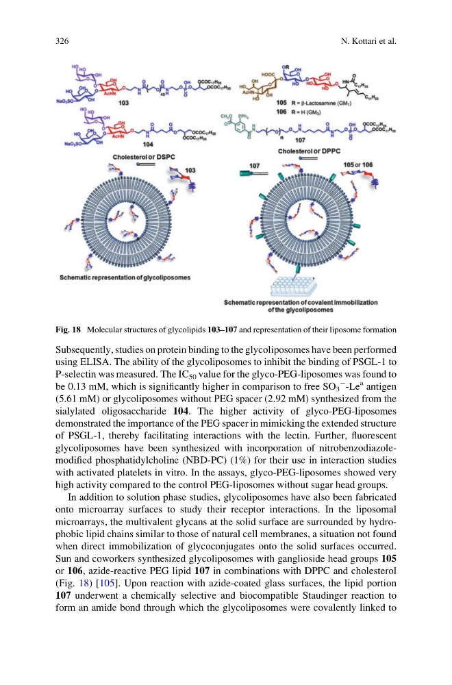
Sun and coworkers synthesized glycoliposomes with ganglioside head groups 105
or 106, azide-reactive PEG lipid 107 in combinations with DPPC and cholesterol
(Fig. 18) [105]. Upon reaction with azide-coated glass surfaces, the lipid portion
107 underwent a chemically selective and biocompatible Staudinger reaction to
form an amide bond through which the glycoliposomes were covalently linked to
Fig. 18 Molecular structures of glycolipids 103–107 and representation of their liposome formation
326
N. Kottari et al.
using ELISA. The ability of the glycoliposomes to inhibit the binding of PSGL-1 to
P-selectin was measured. The IC 50 value for the glyco-PEG-liposomes was found to
be 0.13 mM, which is significantly higher in comparison to free SO 3
À
-Le
a antigen
(5.61 mM) or glycoliposomes without PEG spacer (2.92 mM) synthesized from the
sialylated oligosaccharide 104. The higher activity of glyco-PEG-liposomes
demonstrated the importance of the PEG spacer in mimicking the extended structure
of PSGL-1, thereby facilitating interactions with the lectin. Further, fluorescent
glycoliposomes have been synthesized with incorporation of nitrobenzodiazolemodified phosphatidylcholine (NBD-PC) (1%) for their use in interaction studies
with activated platelets in vitro. In the assays, glyco-PEG-liposomes showed very
high activity compared to the control PEG-liposomes without sugar head groups.
In addition to solution phase studies, glycoliposomes have also been fabricated
onto microarray surfaces to study their receptor interactions. In the liposomal
microarrays, the multivalent glycans at the solid surface are surrounded by hydrophobic lipid chains similar to those of natural cell membranes, a situation not found
when direct immobilization of glycoconjugates onto the solid surfaces occurred.
Sun and coworkers synthesized glycoliposomes with ganglioside head groups 105
or 106, azide-reactive PEG lipid 107 in combinations with DPPC and cholesterol
(Fig. 18) [105]. Upon reaction with azide-coated glass surfaces, the lipid portion
107 underwent a chemically selective and biocompatible Staudinger reaction to
form an amide bond through which the glycoliposomes were covalently linked to
Fig. 18 Molecular structures of glycolipids 103–107 and representation of their liposome formation
326
N. Kottari et al.