

function and a longer stability of the chemical structure of GTP. GTP-loaded PCL/
MWCNT composite nanofibers inhibited cancer cell growth of human hepatoma
cells (Hep G2) and human non-small-cell lung carcinoma cells (A549) but had no
obvious effect on osteoblasts. The A549 cells have an elliptical structure and
conglutinated on nanofiber meshes. Figure 7 displays the fluorescence images and
typical SEM photos of cells cultured for 3 days on GTP-loaded NCF-5 and CF-5
composite nanofiber meshes. In accordance with the toxicity results, they also
observed that the cancer cells were subjected to apoptosis, which could be due to
the released GTP from the nanofibers [150]. A combination of EGCG and caffeic
acid was encapsulated within electrospun PCL nanofibers and its in vitro anticancer
potential evaluated in cultured gastric cancer cell lines (MKN-28). MKN-28 cells
were subjected to apoptosis via activated caspase-3 due to the H 2 O 2 generated by
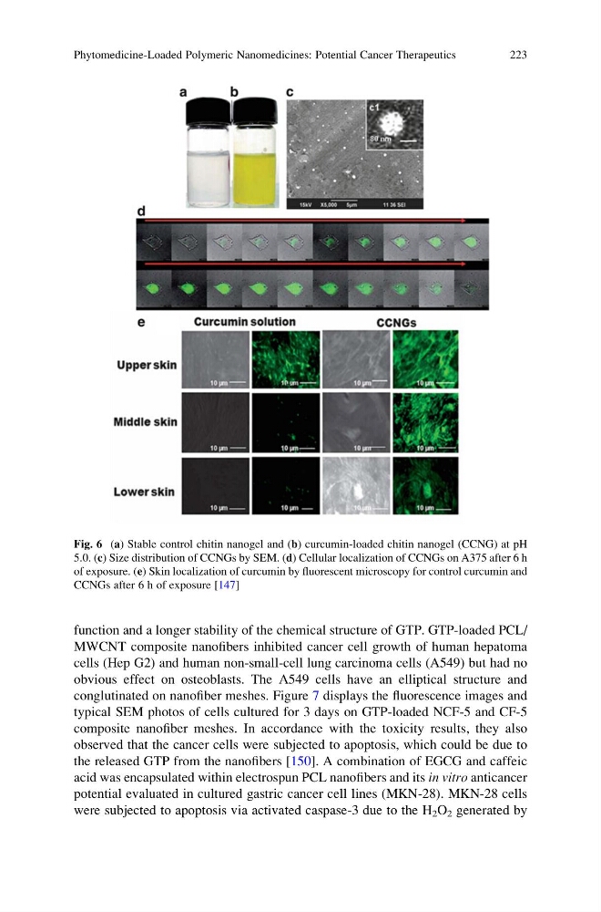
Fig. 6 (a) Stable control chitin nanogel and (b) curcumin-loaded chitin nanogel (CCNG) at pH
5.0. (c) Size distribution of CCNGs by SEM. (d) Cellular localization of CCNGs on A375 after 6 h
of exposure. (e) Skin localization of curcumin by fluorescent microscopy for control curcumin and
CCNGs after 6 h of exposure [147]
Phytomedicine-Loaded Polymeric Nanomedicines: Potential Cancer Therapeutics
223
MWCNT composite nanofibers inhibited cancer cell growth of human hepatoma
cells (Hep G2) and human non-small-cell lung carcinoma cells (A549) but had no
obvious effect on osteoblasts. The A549 cells have an elliptical structure and
conglutinated on nanofiber meshes. Figure 7 displays the fluorescence images and
typical SEM photos of cells cultured for 3 days on GTP-loaded NCF-5 and CF-5
composite nanofiber meshes. In accordance with the toxicity results, they also
observed that the cancer cells were subjected to apoptosis, which could be due to
the released GTP from the nanofibers [150]. A combination of EGCG and caffeic
acid was encapsulated within electrospun PCL nanofibers and its in vitro anticancer
potential evaluated in cultured gastric cancer cell lines (MKN-28). MKN-28 cells
were subjected to apoptosis via activated caspase-3 due to the H 2 O 2 generated by
Fig. 6 (a) Stable control chitin nanogel and (b) curcumin-loaded chitin nanogel (CCNG) at pH
5.0. (c) Size distribution of CCNGs by SEM. (d) Cellular localization of CCNGs on A375 after 6 h
of exposure. (e) Skin localization of curcumin by fluorescent microscopy for control curcumin and
CCNGs after 6 h of exposure [147]
Phytomedicine-Loaded Polymeric Nanomedicines: Potential Cancer Therapeutics
223