

Chitosan-coated cationic poly(butyl)cyanoacrylate (PBCA) nanoparticles
(200 nm) were developed for improving the solubility and bioavailability of
curcumin. Nanocurcumin demonstrated comparable in vitro toxicity and
proapoptotic effects to free curcumin against series of human hepatocellular cancer
cell lines (HepG2, Bel7402, and Huh7 cells). HCC growth in murine xenograft
models was suppressed by the curcumin nanoparticles and also inhibited tumor
angiogenesis by suppressing COX-2 and VEGF expression [135]. Another study
reported that the co-encapsulation of curcumin with doxorubicin in PBCA NPs
(size 133 Æ 5.3 nm) achieved a high reversal efficacy and downregulation
of phosphorylated glycoprotein in an adriamycin-resistant breast cancer cell
line (MCF-7) [136]. Curcumin loaded in crosslinked nanoparticles based on
N-isopropylacrylamide, N-vinyl-2-pyrrolidinone, and PEG acrylate showed more
cytotoxicity in pancreatic cancer cell lines (BxPC3, AsPC1, MiaPaca, XPA-1,
XPA-2, PL-11, PL-12, PL-18, PK-9, and Panc 2.03) and enhanced the bioavailability
(ninefold higher). The authors demonstrated that curcumin nanoparticles were more
active than free curcumin in inhibiting some inflammatory mediators (i.e. TNF-ainduced NF-kB activation) and in suppressing NF-kB-regulated proteins involved
in cell proliferation, invasion, and angiogenesis [137].
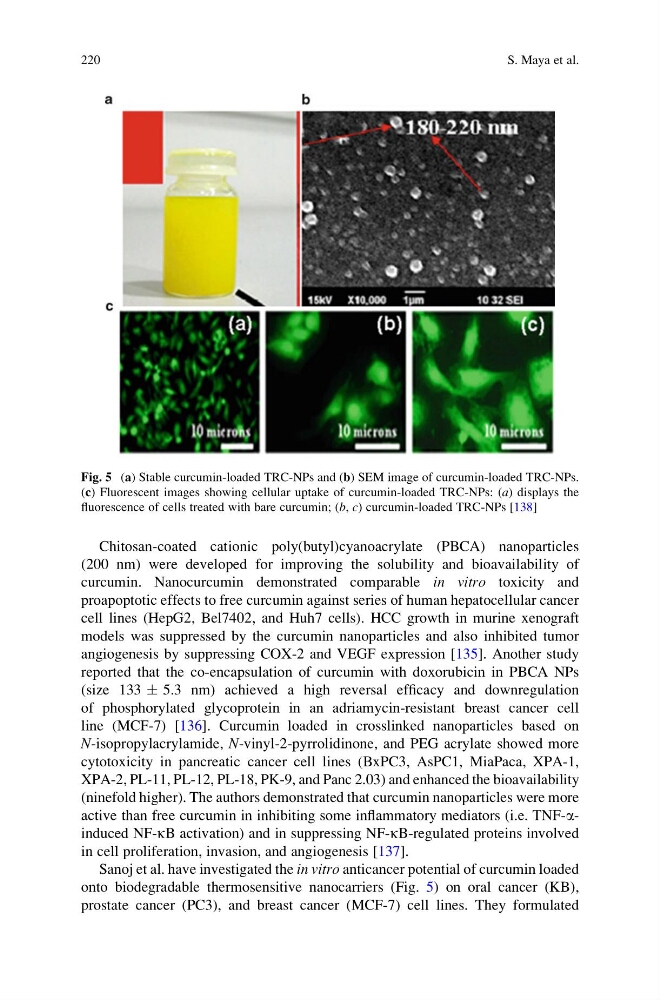
Sanoj et al. have investigated the in vitro anticancer potential of curcumin loaded
onto biodegradable thermosensitive nanocarriers (Fig. 5) on oral cancer (KB),
prostate cancer (PC3), and breast cancer (MCF-7) cell lines. They formulated
Fig. 5 (a) Stable curcumin-loaded TRC-NPs and (b) SEM image of curcumin-loaded TRC-NPs.
(c) Fluorescent images showing cellular uptake of curcumin-loaded TRC-NPs: (a) displays the
fluorescence of cells treated with bare curcumin; (b, c) curcumin-loaded TRC-NPs [138]
220
S. Maya et al.
(200 nm) were developed for improving the solubility and bioavailability of
curcumin. Nanocurcumin demonstrated comparable in vitro toxicity and
proapoptotic effects to free curcumin against series of human hepatocellular cancer
cell lines (HepG2, Bel7402, and Huh7 cells). HCC growth in murine xenograft
models was suppressed by the curcumin nanoparticles and also inhibited tumor
angiogenesis by suppressing COX-2 and VEGF expression [135]. Another study
reported that the co-encapsulation of curcumin with doxorubicin in PBCA NPs
(size 133 Æ 5.3 nm) achieved a high reversal efficacy and downregulation
of phosphorylated glycoprotein in an adriamycin-resistant breast cancer cell
line (MCF-7) [136]. Curcumin loaded in crosslinked nanoparticles based on
N-isopropylacrylamide, N-vinyl-2-pyrrolidinone, and PEG acrylate showed more
cytotoxicity in pancreatic cancer cell lines (BxPC3, AsPC1, MiaPaca, XPA-1,
XPA-2, PL-11, PL-12, PL-18, PK-9, and Panc 2.03) and enhanced the bioavailability
(ninefold higher). The authors demonstrated that curcumin nanoparticles were more
active than free curcumin in inhibiting some inflammatory mediators (i.e. TNF-ainduced NF-kB activation) and in suppressing NF-kB-regulated proteins involved
in cell proliferation, invasion, and angiogenesis [137].
Sanoj et al. have investigated the in vitro anticancer potential of curcumin loaded
onto biodegradable thermosensitive nanocarriers (Fig. 5) on oral cancer (KB),
prostate cancer (PC3), and breast cancer (MCF-7) cell lines. They formulated
Fig. 5 (a) Stable curcumin-loaded TRC-NPs and (b) SEM image of curcumin-loaded TRC-NPs.
(c) Fluorescent images showing cellular uptake of curcumin-loaded TRC-NPs: (a) displays the
fluorescence of cells treated with bare curcumin; (b, c) curcumin-loaded TRC-NPs [138]
220
S. Maya et al.