

of the physical properties including mechanical strength and biological activity.
The rate of degradation of PU depends on the molar mass, degree of crystallinity,
the presence of enzymes, as well as the pH of the medium. PEG is a comonomer
constituent used for the preparation of PUs with excellent properties such as
hydrophilicity, absence of antigenicity, immunogenicity, and nontoxic degradation
products. PUs are frequently used in cell culture, carrier, and biomedical
applications due to their biocompatible and cytocompatible nature. PU shows the
best biocompatibility, durability, and thrombogenic-resistant properties for use in
artificial heart valves [169]. Pentane diisocyanate (PDI)-based PUs are also used in
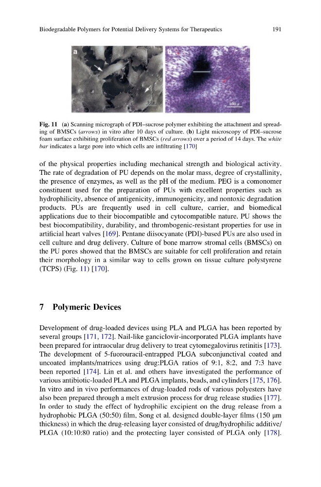
cell culture and drug delivery. Culture of bone marrow stromal cells (BMSCs) on
the PU pores showed that the BMSCs are suitable for cell proliferation and retain
their morphology in a similar way to cells grown on tissue culture polystyrene
(TCPS) (Fig. 11) [170].
7 Polymeric Devices
Development of drug-loaded devices using PLA and PLGA has been reported by
several groups [171, 172]. Nail-like ganciclovir-incorporated PLGA implants have
been prepared for intraocular drug delivery to treat cytomegalovirus retinitis [173].
The development of 5-fuorouracil-entrapped PLGA subconjunctival coated and
uncoated implants/matrices using drug:PLGA ratios of 9:1, 8:2, and 7:3 have
been reported [174]. Lin et al. and others have investigated the performance of
various antibiotic-loaded PLA and PLGA implants, beads, and cylinders [175, 176].
In vitro and in vivo performances of drug-loaded rods of various polyesters have
also been prepared through a melt extrusion process for drug release studies [177].
In order to study the effect of hydrophilic excipient on the drug release from a
hydrophobic PLGA (50:50) film, Song et al. designed double-layer films (150 μm
thickness) in which the drug-releasing layer consisted of drug/hydrophilic additive/
PLGA (10:10:80 ratio) and the protecting layer consisted of PLGA only [178].
Fig. 11 (a) Scanning micrograph of PDI–sucrose polymer exhibiting the attachment and spreading of BMSCs (arrows) in vitro after 10 days of culture. (b) Light microscopy of PDI–sucrose
foam surface exhibiting proliferation of BMSCs (red arrows) over a period of 14 days. The white
bar indicates a large pore into which cells are infiltrating [170]
Biodegradable Polymers for Potential Delivery Systems for Therapeutics
191
The rate of degradation of PU depends on the molar mass, degree of crystallinity,
the presence of enzymes, as well as the pH of the medium. PEG is a comonomer
constituent used for the preparation of PUs with excellent properties such as
hydrophilicity, absence of antigenicity, immunogenicity, and nontoxic degradation
products. PUs are frequently used in cell culture, carrier, and biomedical
applications due to their biocompatible and cytocompatible nature. PU shows the
best biocompatibility, durability, and thrombogenic-resistant properties for use in
artificial heart valves [169]. Pentane diisocyanate (PDI)-based PUs are also used in
cell culture and drug delivery. Culture of bone marrow stromal cells (BMSCs) on
the PU pores showed that the BMSCs are suitable for cell proliferation and retain
their morphology in a similar way to cells grown on tissue culture polystyrene
(TCPS) (Fig. 11) [170].
7 Polymeric Devices
Development of drug-loaded devices using PLA and PLGA has been reported by
several groups [171, 172]. Nail-like ganciclovir-incorporated PLGA implants have
been prepared for intraocular drug delivery to treat cytomegalovirus retinitis [173].
The development of 5-fuorouracil-entrapped PLGA subconjunctival coated and
uncoated implants/matrices using drug:PLGA ratios of 9:1, 8:2, and 7:3 have
been reported [174]. Lin et al. and others have investigated the performance of
various antibiotic-loaded PLA and PLGA implants, beads, and cylinders [175, 176].
In vitro and in vivo performances of drug-loaded rods of various polyesters have
also been prepared through a melt extrusion process for drug release studies [177].
In order to study the effect of hydrophilic excipient on the drug release from a
hydrophobic PLGA (50:50) film, Song et al. designed double-layer films (150 μm
thickness) in which the drug-releasing layer consisted of drug/hydrophilic additive/
PLGA (10:10:80 ratio) and the protecting layer consisted of PLGA only [178].
Fig. 11 (a) Scanning micrograph of PDI–sucrose polymer exhibiting the attachment and spreading of BMSCs (arrows) in vitro after 10 days of culture. (b) Light microscopy of PDI–sucrose
foam surface exhibiting proliferation of BMSCs (red arrows) over a period of 14 days. The white
bar indicates a large pore into which cells are infiltrating [170]
Biodegradable Polymers for Potential Delivery Systems for Therapeutics
191