

present in the bone matrix are induced to resorb old bone. Osteoblasts are
stimulated by insulin-like growth factors I and II to deposit calcium-containing
minerals secreted by osteoclasts and/or osteocytes. New bone formation is
controlled by osteocytes by modulation of osteoblast differentiation from noncalcium-depositing cells to calcium-depositing cells through secretion of growth
factors such as insulin-like growth factor-I and tissue growth factor β [27].
Bone, like other connective tissue in the embryo, is derived from mesenchymal
cells. These cells have the ability to proliferate and differentiate into bone cells,
which are known as osteoprogenitor cells. They are also called bone-precursor cells.
2.4.1 Osteoblasts
Osteoblast cells, found on the surface of periosteal and endosteal surfaces, are
responsible for the formation of new bone tissue. They has an average diameter
of 10–50 μm and produce a matrix of osteoid, which is composed mainly of type
I collagen. Osteoblasts are also responsible for mineralization of this matrix.
Osteoblasts synthesize and deposit bone sialoprotein, osteocalcin (a calciumbinding protein), and other matrix proteins at the beginning of the mineralization
process. Osteocalcin interacts with HAp and is thought to mediate pairing to
bone resorption by osteoclasts and bone formation by osteoblasts and/or
osteocytes.
4800 μm
350 μm
200 μm 40 μm
SIDE
VIEW
REMODELING
SPEED=
40μm/day
OSTEOBLASTS
OSTEOCLASTS
CROSS-SECTIONAL
VIEWS
VASCULATURE
a
b
c
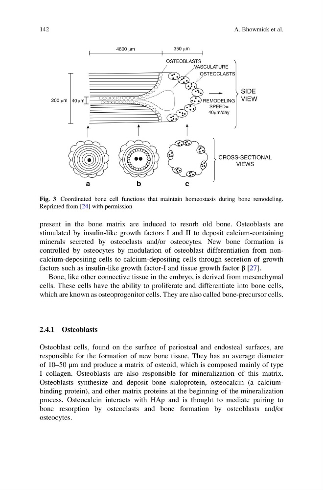
Fig. 3 Coordinated bone cell functions that maintain homeostasis during bone remodeling.
Reprinted from [24] with permission
142
A. Bhowmick et al.
stimulated by insulin-like growth factors I and II to deposit calcium-containing
minerals secreted by osteoclasts and/or osteocytes. New bone formation is
controlled by osteocytes by modulation of osteoblast differentiation from noncalcium-depositing cells to calcium-depositing cells through secretion of growth
factors such as insulin-like growth factor-I and tissue growth factor β [27].
Bone, like other connective tissue in the embryo, is derived from mesenchymal
cells. These cells have the ability to proliferate and differentiate into bone cells,
which are known as osteoprogenitor cells. They are also called bone-precursor cells.
2.4.1 Osteoblasts
Osteoblast cells, found on the surface of periosteal and endosteal surfaces, are
responsible for the formation of new bone tissue. They has an average diameter
of 10–50 μm and produce a matrix of osteoid, which is composed mainly of type
I collagen. Osteoblasts are also responsible for mineralization of this matrix.
Osteoblasts synthesize and deposit bone sialoprotein, osteocalcin (a calciumbinding protein), and other matrix proteins at the beginning of the mineralization
process. Osteocalcin interacts with HAp and is thought to mediate pairing to
bone resorption by osteoclasts and bone formation by osteoblasts and/or
osteocytes.
4800 μm
350 μm
200 μm 40 μm
SIDE
VIEW
REMODELING
SPEED=
40μm/day
OSTEOBLASTS
OSTEOCLASTS
CROSS-SECTIONAL
VIEWS
VASCULATURE
a
b
c
Fig. 3 Coordinated bone cell functions that maintain homeostasis during bone remodeling.
Reprinted from [24] with permission
142
A. Bhowmick et al.