

rigidity to ensure that the growing array does not bend back on itself, thereby
poisoning the growth of the lattice. Figure 8 shows three lattices [10, 11] built from
motifs shown in Fig. 7. Figure 8a shows a two-tile array that alternates DX motifs
with DX + J motifs. The extra domain in the DX + J motif leads to a stripe in the
pattern. The size of the motifs is 16 nm in the horizontal direction, so the stripes
should be separated by 32 nm, which can be seen in the atomic force micrograph on
the right. A related array is shown in Fig. 8b, where three DX motifs and a single
DX + J motif are seen to form an array with ~64 nm stripes. Figure 8c illustrates a
motif made from two TX motifs connected from the top of one to the bottom of the
other (A and B), creating gaps in the lattice. The gaps are filled by a rotated TX
molecule (C’) and by a duplex (D). The AFM image can be seen on the right.
The success in self-assembling the variety of 2D arrays shown in Fig. 8 suggests
that it ought to be possible to organize DNA motifs into 3D crystals. The criteria for
evaluating crystals are stricter than those for evaluating 2D arrays: Atomic force
micrographs usually yield resolutions of about 7–10 nm, but crystals of DNA must
diffract X-rays to at least 4–5 A ˚ to be readily interpretable. The motif that has been
used to produce 3D crystals is the tensegrity triangle [12], first developed by
Chengde Mao. Sticky ends can be added to these molecules to produce selfassembled designed rhombohedral crystals of the requisite resolution [13]. Figure 9a
illustrates the environment of a single tensegrity triangle in a crystal. The three
helices are colored differently, and it is evident that the axes of the three helices
point in three independent directions in space. Figure 9b shows that the centers of
the triangles can be placed on the vertices of a rhombohedron. The cavity within the
rhombohedron has a volume of about 100 nm
3 .
A large variety of nanomechanical devices have been produced from DNA. The
most interesting ones are those that avail themselves of the programmability of
DNA because they can be individually addressed, thereby enabling their states to be
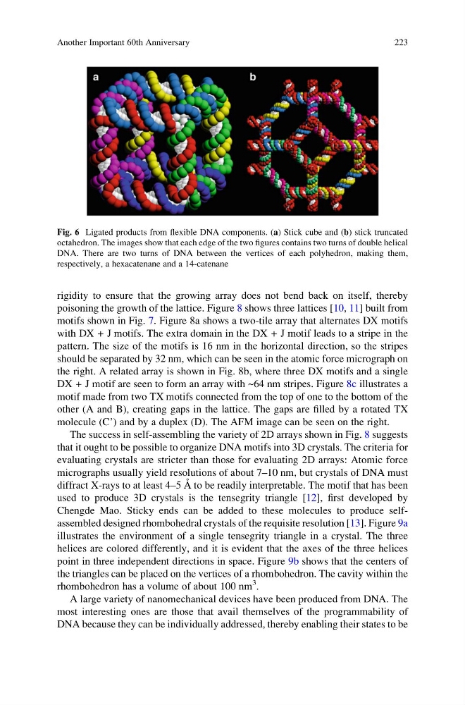
Fig. 6 Ligated products from flexible DNA components. (a) Stick cube and (b) stick truncated
octahedron. The images show that each edge of the two figures contains two turns of double helical
DNA. There are two turns of DNA between the vertices of each polyhedron, making them,
respectively, a hexacatenane and a 14-catenane
Another Important 60th Anniversary
223
poisoning the growth of the lattice. Figure 8 shows three lattices [10, 11] built from
motifs shown in Fig. 7. Figure 8a shows a two-tile array that alternates DX motifs
with DX + J motifs. The extra domain in the DX + J motif leads to a stripe in the
pattern. The size of the motifs is 16 nm in the horizontal direction, so the stripes
should be separated by 32 nm, which can be seen in the atomic force micrograph on
the right. A related array is shown in Fig. 8b, where three DX motifs and a single
DX + J motif are seen to form an array with ~64 nm stripes. Figure 8c illustrates a
motif made from two TX motifs connected from the top of one to the bottom of the
other (A and B), creating gaps in the lattice. The gaps are filled by a rotated TX
molecule (C’) and by a duplex (D). The AFM image can be seen on the right.
The success in self-assembling the variety of 2D arrays shown in Fig. 8 suggests
that it ought to be possible to organize DNA motifs into 3D crystals. The criteria for
evaluating crystals are stricter than those for evaluating 2D arrays: Atomic force
micrographs usually yield resolutions of about 7–10 nm, but crystals of DNA must
diffract X-rays to at least 4–5 A ˚ to be readily interpretable. The motif that has been
used to produce 3D crystals is the tensegrity triangle [12], first developed by
Chengde Mao. Sticky ends can be added to these molecules to produce selfassembled designed rhombohedral crystals of the requisite resolution [13]. Figure 9a
illustrates the environment of a single tensegrity triangle in a crystal. The three
helices are colored differently, and it is evident that the axes of the three helices
point in three independent directions in space. Figure 9b shows that the centers of
the triangles can be placed on the vertices of a rhombohedron. The cavity within the
rhombohedron has a volume of about 100 nm
3 .
A large variety of nanomechanical devices have been produced from DNA. The
most interesting ones are those that avail themselves of the programmability of
DNA because they can be individually addressed, thereby enabling their states to be
Fig. 6 Ligated products from flexible DNA components. (a) Stick cube and (b) stick truncated
octahedron. The images show that each edge of the two figures contains two turns of double helical
DNA. There are two turns of DNA between the vertices of each polyhedron, making them,
respectively, a hexacatenane and a 14-catenane
Another Important 60th Anniversary
223