

cellular proteins by affinity chromatography, and identified by high-throughput
mass spectrometry. Because other cellular proteins are essentially inert to the
tagging chemistry, proteins made during the Aha pulse are highly enriched and
easily identified. Tagging with dyes allows one to determine the cellular locations
of the proteins made during the Aha pulse (Fig. 5) [37].
3.2 Cell-Selective BONCAT
In studies of complex biological systems, it is often important to determine what is
going on in one particular type of cell, rather than averaging information obtained
from many different cell types. Labeling with Aha does not discriminate among
cells because Aha is activated for protein synthesis by the wild-type methionyltRNA synthetase (MetRS) present in all cells. To enable cell-selective labeling, we
developed the longer-chain methionine analog azidonorleucine (Anl), which is not
a good substrate for the wild-type MetRS and requires a mutant synthetase for
activation. Cell-selective labeling can then be achieved by ensuring that the mutant
MetRS is expressed only in the cells of interest.
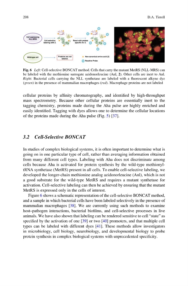
Figure 6 shows a schematic representation of the cell-selective BONCAT method,
and a sample in which bacterial cells have been labeled selectively in the presence of
mammalian macrophages [38]. We are currently using such methods to examine
host–pathogen interactions, bacterial biofilms, and cell-selective processes in live
animals. We have also shown that labeling can be rendered sensitive to cell “state” as
specified by the activation of one [39] or two [40] promoters, and that multiple cell
types can be labeled with different dyes [41]. These methods allow investigators
in microbiology, cell biology, neurobiology, and developmental biology to probe
protein synthesis in complex biological systems with unprecedented specificity.
Fig. 6 Left: Cell-selective BONCAT method. Cells that carry the mutant MetRS (NLL-MRS) can
be labeled with the methionine surrogate azidonorleucine (Anl, 2). Other cells are inert to Anl.
Right: Bacterial cells carrying the NLL synthetase are labeled with a fluorescent alkyne dye
(green) in the presence of mammalian macrophages (red). Macrophage proteins are not labeled
208
D.A. Tirrell
mass spectrometry. Because other cellular proteins are essentially inert to the
tagging chemistry, proteins made during the Aha pulse are highly enriched and
easily identified. Tagging with dyes allows one to determine the cellular locations
of the proteins made during the Aha pulse (Fig. 5) [37].
3.2 Cell-Selective BONCAT
In studies of complex biological systems, it is often important to determine what is
going on in one particular type of cell, rather than averaging information obtained
from many different cell types. Labeling with Aha does not discriminate among
cells because Aha is activated for protein synthesis by the wild-type methionyltRNA synthetase (MetRS) present in all cells. To enable cell-selective labeling, we
developed the longer-chain methionine analog azidonorleucine (Anl), which is not
a good substrate for the wild-type MetRS and requires a mutant synthetase for
activation. Cell-selective labeling can then be achieved by ensuring that the mutant
MetRS is expressed only in the cells of interest.
Figure 6 shows a schematic representation of the cell-selective BONCAT method,
and a sample in which bacterial cells have been labeled selectively in the presence of
mammalian macrophages [38]. We are currently using such methods to examine
host–pathogen interactions, bacterial biofilms, and cell-selective processes in live
animals. We have also shown that labeling can be rendered sensitive to cell “state” as
specified by the activation of one [39] or two [40] promoters, and that multiple cell
types can be labeled with different dyes [41]. These methods allow investigators
in microbiology, cell biology, neurobiology, and developmental biology to probe
protein synthesis in complex biological systems with unprecedented specificity.
Fig. 6 Left: Cell-selective BONCAT method. Cells that carry the mutant MetRS (NLL-MRS) can
be labeled with the methionine surrogate azidonorleucine (Anl, 2). Other cells are inert to Anl.
Right: Bacterial cells carrying the NLL synthetase are labeled with a fluorescent alkyne dye
(green) in the presence of mammalian macrophages (red). Macrophage proteins are not labeled
208
D.A. Tirrell