

enhance blood circulation, shield epitopes at the protein surface, reduce proteolysis by
proteases, and enable drug uptake into the lipophilic interior of albumin.
However, if substantially higher drug loading is required, both the covalent
attachment of drugs to the globular HSA protein as well as the non-covalent adsorption into the binding pockets are limited by the available space. In order to further
increase the drug loading capacity of HSA, reactive groups within the scaffold of
HSA that are normally hidden inside the protein scaffold need to become more
accessible for chemical modifications.
1.4 Albumin Copolymer Polyelectrolytes Allow
Efficient Drug Delivery
Further exploration of multifunctional protein polyelectrolytes requires the full use of
the inner functional groups. However, unfolding of a protein’s tertiary structure often
leads to precipitation and destabilization. Recently, an in situ stabilization method has
been developed that allows the unfolding of proteins into polypeptide chains and
exposure of “hidden” functional groups. This has been achieved by controlled
chemical denaturation of the native proteins and an in situ stabilization with PEO
chains (Fig. 6a). Different protein precursors (e.g., HSA, BSA, and the protease
lysozyme) have been studied to prove the general applicability of this approach
[41, 42]. The protein-derived polypeptide side chain copolymers feature precisely
defined backbone lengths, large numbers of readily available functional groups, and
partially maintained secondary structure elements [41, 42]. Modification of the
exposed functional groups allows successful conjugation onto the protein backbone
of multiple reactive moieties such as chromophores, anticancer drugs, or MRI
contrast agents [43, 44]. Due to the presence of hydrophobic patches along the protein
primary sequence, the unfolded albumin-based polypeptides can efficiently interact
with hydrophobic molecules. Polymeric micelles are formed where the hydrophilic
groups face to the outside, and hydrophobic drug molecules are encapsulated inside
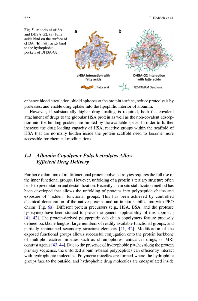
Fig. 5 Models of cHSA
and DHSA-G2. (a) Fatty
acids bind on the surface of
cHSA. (b) Fatty acids bind
to the hydrophobic
pockets of DHSA-G2
222
J. Hedrich et al.
proteases, and enable drug uptake into the lipophilic interior of albumin.
However, if substantially higher drug loading is required, both the covalent
attachment of drugs to the globular HSA protein as well as the non-covalent adsorption into the binding pockets are limited by the available space. In order to further
increase the drug loading capacity of HSA, reactive groups within the scaffold of
HSA that are normally hidden inside the protein scaffold need to become more
accessible for chemical modifications.
1.4 Albumin Copolymer Polyelectrolytes Allow
Efficient Drug Delivery
Further exploration of multifunctional protein polyelectrolytes requires the full use of
the inner functional groups. However, unfolding of a protein’s tertiary structure often
leads to precipitation and destabilization. Recently, an in situ stabilization method has
been developed that allows the unfolding of proteins into polypeptide chains and
exposure of “hidden” functional groups. This has been achieved by controlled
chemical denaturation of the native proteins and an in situ stabilization with PEO
chains (Fig. 6a). Different protein precursors (e.g., HSA, BSA, and the protease
lysozyme) have been studied to prove the general applicability of this approach
[41, 42]. The protein-derived polypeptide side chain copolymers feature precisely
defined backbone lengths, large numbers of readily available functional groups, and
partially maintained secondary structure elements [41, 42]. Modification of the
exposed functional groups allows successful conjugation onto the protein backbone
of multiple reactive moieties such as chromophores, anticancer drugs, or MRI
contrast agents [43, 44]. Due to the presence of hydrophobic patches along the protein
primary sequence, the unfolded albumin-based polypeptides can efficiently interact
with hydrophobic molecules. Polymeric micelles are formed where the hydrophilic
groups face to the outside, and hydrophobic drug molecules are encapsulated inside
Fig. 5 Models of cHSA
and DHSA-G2. (a) Fatty
acids bind on the surface of
cHSA. (b) Fatty acids bind
to the hydrophobic
pockets of DHSA-G2
222
J. Hedrich et al.