

Mixtures of lysozyme and succinylated lysozyme are not the only
protein–protein systems that have been studied. Nigen et al. studied the formation
of microspheres of calcium-depleted a-lactalbumin and lysozyme at 45
C and
pH 7.5. These microspheres contain equimolar ratios of the two protein molecules.
With confocal scanning laser microscopy (CSLM), and the two protein molecules
labelled in different colours, the authors showed that the microspheres are built-up
by binary assemblies of lysozyme and a-lactalbumin [89].
The ionic strength is important for the formation of a-lactalbumin–lysozyme
microspheres. The equilibrium between free protein molecules and protein molecules
in the microspheres depends on the ionic strength, as also found for the lysozyme–
succinylated lysozyme system [82]. Increasing the salt concentration decreases the
size of the microspheres; above 124 mM salt no microspheres are formed. Addition
of salt to existing microspheres decreases the number of protein molecules within the
spheres, but the protein aggregates remain spherical and monodisperse [90].
The protein molecules are able to exchange between different spheres. This was
studied by CSLM using two different coloured batches (fluorescent green and red)
of lysozyme molecules. Red fluorescent lysozyme molecules were added to
1000
C
salt (mM)
100
10
1
0.0001
0.001
0.01
0.1
1
increasing T
12
x=14
10
2f
f 0
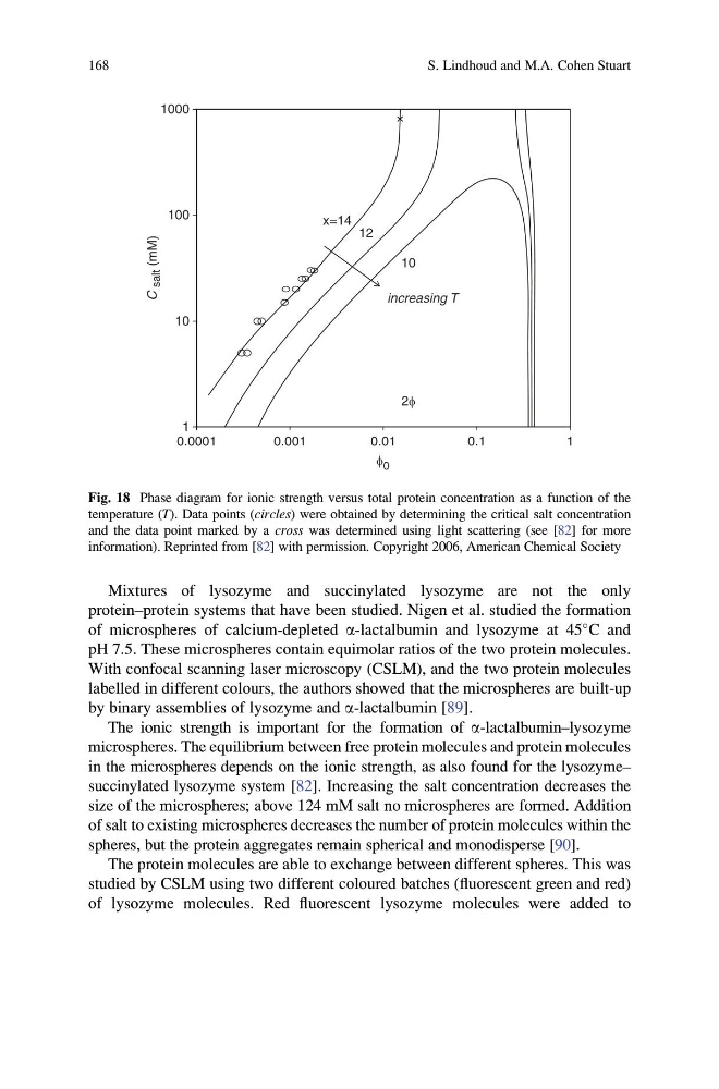
Fig. 18 Phase diagram for ionic strength versus total protein concentration as a function of the
temperature (T). Data points (circles) were obtained by determining the critical salt concentration
and the data point marked by a cross was determined using light scattering (see [82] for more
information). Reprinted from [82] with permission. Copyright 2006, American Chemical Society
168
S. Lindhoud and M.A. Cohen Stuart
protein–protein systems that have been studied. Nigen et al. studied the formation
of microspheres of calcium-depleted a-lactalbumin and lysozyme at 45
C and
pH 7.5. These microspheres contain equimolar ratios of the two protein molecules.
With confocal scanning laser microscopy (CSLM), and the two protein molecules
labelled in different colours, the authors showed that the microspheres are built-up
by binary assemblies of lysozyme and a-lactalbumin [89].
The ionic strength is important for the formation of a-lactalbumin–lysozyme
microspheres. The equilibrium between free protein molecules and protein molecules
in the microspheres depends on the ionic strength, as also found for the lysozyme–
succinylated lysozyme system [82]. Increasing the salt concentration decreases the
size of the microspheres; above 124 mM salt no microspheres are formed. Addition
of salt to existing microspheres decreases the number of protein molecules within the
spheres, but the protein aggregates remain spherical and monodisperse [90].
The protein molecules are able to exchange between different spheres. This was
studied by CSLM using two different coloured batches (fluorescent green and red)
of lysozyme molecules. Red fluorescent lysozyme molecules were added to
1000
C
salt (mM)
100
10
1
0.0001
0.001
0.01
0.1
1
increasing T
12
x=14
10
2f
f 0
Fig. 18 Phase diagram for ionic strength versus total protein concentration as a function of the
temperature (T). Data points (circles) were obtained by determining the critical salt concentration
and the data point marked by a cross was determined using light scattering (see [82] for more
information). Reprinted from [82] with permission. Copyright 2006, American Chemical Society
168
S. Lindhoud and M.A. Cohen Stuart