

Using the whisker method introduced by Ihn et al. [47], Guillerez and coworkers
followed the formation of P3HT nanofibrils in solution, prepared using a marginal
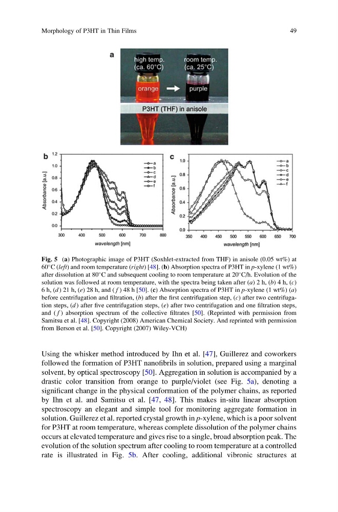
solvent, by optical spectroscopy [50]. Aggregation in solution is accompanied by a
drastic color transition from orange to purple/violet (see Fig. 5a), denoting a
significant change in the physical conformation of the polymer chains, as reported
by Ihn et al. and Samitsu et al. [47, 48]. This makes in-situ linear absorption
spectroscopy an elegant and simple tool for monitoring aggregate formation in
solution. Guillerez et al. reported crystal growth in p-xylene, which is a poor solvent
for P3HT at room temperature, whereas complete dissolution of the polymer chains
occurs at elevated temperature and gives rise to a single, broad absorption peak. The
evolution of the solution spectrum after cooling to room temperature at a controlled
rate is illustrated in Fig. 5b. After cooling, additional vibronic structures at
Fig. 5 (a) Photographic image of P3HT (Soxhlet-extracted from THF) in anisole (0.05 wt%) at
60
C (left) and room temperature (right) [48]. (b) Absorption spectra of P3HT in p-xylene (1 wt%)
after dissolution at 80
C and subsequent cooling to room temperature at 20
C/h. Evolution of the
solution was followed at room temperature, with the spectra being taken after (a) 2 h, (b) 4 h, (c)
6 h, (d ) 21 h, (e) 28 h, and ( f ) 48 h [50]. (c) Absorption spectra of P3HT in p-xylene (1 wt%) (a)
before centrifugation and filtration, (b) after the first centrifugation step, (c) after two centrifugation steps, (d ) after five centrifugation steps, (e) after two centrifugation and one filtration steps,
and ( f ) absorption spectrum of the collective filtrates [50]. (Reprinted with permission from
Samitsu et al. [48]. Copyright (2008) American Chemical Society. And reprinted with permission
from Berson et al. [50]. Copyright (2007) Wiley-VCH)
Morphology of P3HT in Thin Films
49
followed the formation of P3HT nanofibrils in solution, prepared using a marginal
solvent, by optical spectroscopy [50]. Aggregation in solution is accompanied by a
drastic color transition from orange to purple/violet (see Fig. 5a), denoting a
significant change in the physical conformation of the polymer chains, as reported
by Ihn et al. and Samitsu et al. [47, 48]. This makes in-situ linear absorption
spectroscopy an elegant and simple tool for monitoring aggregate formation in
solution. Guillerez et al. reported crystal growth in p-xylene, which is a poor solvent
for P3HT at room temperature, whereas complete dissolution of the polymer chains
occurs at elevated temperature and gives rise to a single, broad absorption peak. The
evolution of the solution spectrum after cooling to room temperature at a controlled
rate is illustrated in Fig. 5b. After cooling, additional vibronic structures at
Fig. 5 (a) Photographic image of P3HT (Soxhlet-extracted from THF) in anisole (0.05 wt%) at
60
C (left) and room temperature (right) [48]. (b) Absorption spectra of P3HT in p-xylene (1 wt%)
after dissolution at 80
C and subsequent cooling to room temperature at 20
C/h. Evolution of the
solution was followed at room temperature, with the spectra being taken after (a) 2 h, (b) 4 h, (c)
6 h, (d ) 21 h, (e) 28 h, and ( f ) 48 h [50]. (c) Absorption spectra of P3HT in p-xylene (1 wt%) (a)
before centrifugation and filtration, (b) after the first centrifugation step, (c) after two centrifugation steps, (d ) after five centrifugation steps, (e) after two centrifugation and one filtration steps,
and ( f ) absorption spectrum of the collective filtrates [50]. (Reprinted with permission from
Samitsu et al. [48]. Copyright (2008) American Chemical Society. And reprinted with permission
from Berson et al. [50]. Copyright (2007) Wiley-VCH)
Morphology of P3HT in Thin Films
49