

molecular weight of 29 kg/mol, exceeds the width of the nanofibrils suggests that
the chains fold back and forth in order to be incorporated into the nanostructures, a
typical feature of semicrystalline polymers. Zhai and coworkers could further show
that the fibril width increases with molecular weight, but saturates above a critical
value of around 10 kg/mol (M n ) [49]. Below the critical molecular weight,
nanofibrils are assumed to be formed by extended chains that span the width of
the fibrils. Above the critical molecular weight, chain folding takes place and leads
to a saturation of fibril width. The fold-length is dependent on the crystallization
temperature in solution, which is characteristic of the crystallization of polymers.
Since the seminal work of Ihn et al. in 1993, a large number of publications have
followed that deal with the structural and optical characterization of nanofibrils, but
also address their performance in devices such as organic field-effect transistors
(OFETs) and organic solar cells (OSCs) [47, 48, 50–53]. In general, two common
approaches for P3AT nanofibril production are described in the literature [52]:
(1) the whisker method and (2) the mixed solvent method. For both approaches, the
growth of 1D nanocrystals is driven by strong π–π-interactions and unfavorable
interactions between the solvent and polymer backbone under limited solubility.
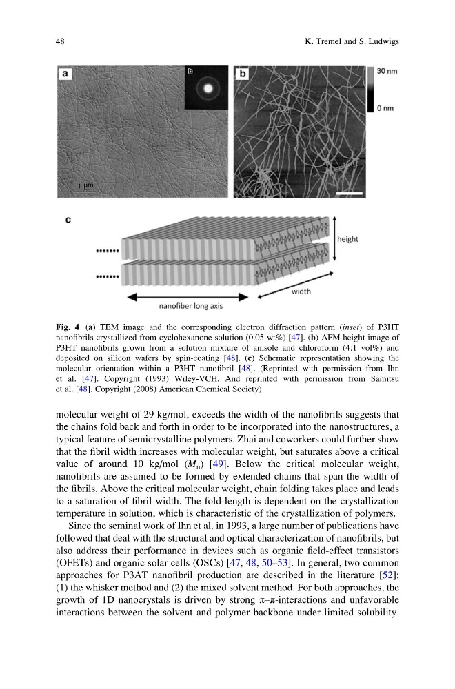
Fig. 4 (a) TEM image and the corresponding electron diffraction pattern (inset) of P3HT
nanofibrils crystallized from cyclohexanone solution (0.05 wt%) [47]. (b) AFM height image of
P3HT nanofibrils grown from a solution mixture of anisole and chloroform (4:1 vol%) and
deposited on silicon wafers by spin-coating [48]. (c) Schematic representation showing the
molecular orientation within a P3HT nanofibril [48]. (Reprinted with permission from Ihn
et al. [47]. Copyright (1993) Wiley-VCH. And reprinted with permission from Samitsu
et al. [48]. Copyright (2008) American Chemical Society)
48
K. Tremel and S. Ludwigs
the chains fold back and forth in order to be incorporated into the nanostructures, a
typical feature of semicrystalline polymers. Zhai and coworkers could further show
that the fibril width increases with molecular weight, but saturates above a critical
value of around 10 kg/mol (M n ) [49]. Below the critical molecular weight,
nanofibrils are assumed to be formed by extended chains that span the width of
the fibrils. Above the critical molecular weight, chain folding takes place and leads
to a saturation of fibril width. The fold-length is dependent on the crystallization
temperature in solution, which is characteristic of the crystallization of polymers.
Since the seminal work of Ihn et al. in 1993, a large number of publications have
followed that deal with the structural and optical characterization of nanofibrils, but
also address their performance in devices such as organic field-effect transistors
(OFETs) and organic solar cells (OSCs) [47, 48, 50–53]. In general, two common
approaches for P3AT nanofibril production are described in the literature [52]:
(1) the whisker method and (2) the mixed solvent method. For both approaches, the
growth of 1D nanocrystals is driven by strong π–π-interactions and unfavorable
interactions between the solvent and polymer backbone under limited solubility.
Fig. 4 (a) TEM image and the corresponding electron diffraction pattern (inset) of P3HT
nanofibrils crystallized from cyclohexanone solution (0.05 wt%) [47]. (b) AFM height image of
P3HT nanofibrils grown from a solution mixture of anisole and chloroform (4:1 vol%) and
deposited on silicon wafers by spin-coating [48]. (c) Schematic representation showing the
molecular orientation within a P3HT nanofibril [48]. (Reprinted with permission from Ihn
et al. [47]. Copyright (1993) Wiley-VCH. And reprinted with permission from Samitsu
et al. [48]. Copyright (2008) American Chemical Society)
48
K. Tremel and S. Ludwigs