

The tertiary structure of the proteins is the 3D organisation of the proteins,
involving the repartition of the secondary structure units of the proteins with respect
to each other. The driving force for the folding into tertiary structure is dependent on
the physicochemical properties of the medium. In aqueous solutions, the hydrophobic residues of the proteins are hidden in the core of the proteins in order to
minimise contacts with water molecules. The protein tertiary structure is stabilised
by hydrophobic interactions, salt bridges, hydrogen bonds and, for some proteins,
disulfide bonds.
Behind these three structural levels, some proteins have a quaternary structure
that results from the association of two or more identical or different polypeptide
chains (protein subunits).
In solutions, the overall protein conformation fluctuates between a large number
of conformations that are very similar to each other. The native protein structure is
defined as the structure of lowest energy or structure of highest probability. Away
from their pI, proteins in solution are stable because the protein molecules carry
charges of the same sign and repel each other. In contrast to intrinsically unfolded
proteins, numerous globular proteins are also stable close to their pI at low or
medium ionic strength due to the presence of residual charged patches on the
protein surface that counterbalance short-range attractive interactions. In some
conditions (elevated temperatures, presence of denaturants, etc.), the native protein
unfolds (loses its native conformation) or denatures. Each protein (except intrinsically unfolded proteins) has a denaturation temperature that is dependent on the
conditions of the medium (pH, ionic strength, dielectric constant). Upon denaturation, the hydrodynamic size, the flexibility and also the reactivity of the proteins
increase because of the exposure of reactive groups to the surface of the proteins.
-35
-30
-25
-20
-15
-10
-5
0
5
10
15
20
1
2
3
4
5
6
7
8
9
10
11
12
pH
net charge
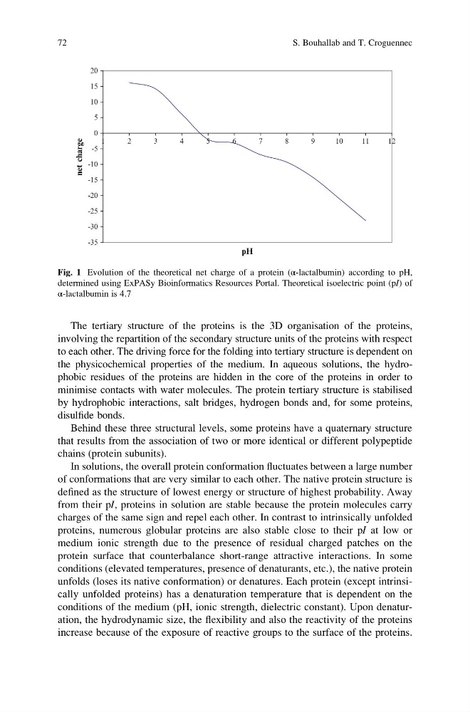
Fig. 1 Evolution of the theoretical net charge of a protein (α-lactalbumin) according to pH,
determined using ExPASy Bioinformatics Resources Portal. Theoretical isoelectric point (pI) of
α-lactalbumin is 4.7
72
S. Bouhallab and T. Croguennec
involving the repartition of the secondary structure units of the proteins with respect
to each other. The driving force for the folding into tertiary structure is dependent on
the physicochemical properties of the medium. In aqueous solutions, the hydrophobic residues of the proteins are hidden in the core of the proteins in order to
minimise contacts with water molecules. The protein tertiary structure is stabilised
by hydrophobic interactions, salt bridges, hydrogen bonds and, for some proteins,
disulfide bonds.
Behind these three structural levels, some proteins have a quaternary structure
that results from the association of two or more identical or different polypeptide
chains (protein subunits).
In solutions, the overall protein conformation fluctuates between a large number
of conformations that are very similar to each other. The native protein structure is
defined as the structure of lowest energy or structure of highest probability. Away
from their pI, proteins in solution are stable because the protein molecules carry
charges of the same sign and repel each other. In contrast to intrinsically unfolded
proteins, numerous globular proteins are also stable close to their pI at low or
medium ionic strength due to the presence of residual charged patches on the
protein surface that counterbalance short-range attractive interactions. In some
conditions (elevated temperatures, presence of denaturants, etc.), the native protein
unfolds (loses its native conformation) or denatures. Each protein (except intrinsically unfolded proteins) has a denaturation temperature that is dependent on the
conditions of the medium (pH, ionic strength, dielectric constant). Upon denaturation, the hydrodynamic size, the flexibility and also the reactivity of the proteins
increase because of the exposure of reactive groups to the surface of the proteins.
-35
-30
-25
-20
-15
-10
-5
0
5
10
15
20
1
2
3
4
5
6
7
8
9
10
11
12
pH
net charge
Fig. 1 Evolution of the theoretical net charge of a protein (α-lactalbumin) according to pH,
determined using ExPASy Bioinformatics Resources Portal. Theoretical isoelectric point (pI) of
α-lactalbumin is 4.7
72
S. Bouhallab and T. Croguennec