

polyelectrolytes whose charge sign and density show a strong dependence on the
pH. Proteins are distinguished from each other by their amino acid sequence, their
folding (globular folded form, fibrous form, intrinsically unfolded conformation)
and their biological function. One of the key features of proteins is their ability to
acquire distinctive secondary and three-dimensional (3D) structures dictated by the
amino acid sequence.
The primary structure of a protein is the succession of amino acids bonded
together by peptide bonds between the carboxyl group of the amino acid N and the
amine group of the amino acid N + 1. The acid hydrolysis of proteins releases 20
amino acids, each having a lateral chain with specific reactivity. Some lateral chains
are hydrophobic, others are hydrophilic, neutral or carry an electric charge (positive
or negative). The presence of hydrophobic residues confers to proteins an affinity
towards hydrophobic surfaces. The charge on the lateral chain of the amino acids
gives the protein a net charge that is positive, negative or null according to the pH
value. At a certain pH, referred to as the isoelectric point (pI), the numbers of positive
and negative charges on a protein are equal and the protein is electrically neutral.
Proteins have an excess of positive charges below its pI and an excess of negative
charges above its pI. Basic proteins (high pI) are rich in arginine and lysine residues
whereas acidic proteins are rich in glutamic and aspartic acid residues. Figure 1
illustrates an example of how the theoretical net charge of a globular protein (α-La)
evolves according to pH, with a zero net electric charge (pI) around 4.6.
The secondary structure consists of the local spatial arrangement of the polypeptide chain into repeating structures stabilised by hydrogen bonds, i.e. α-helices and
β-sheets. In α-helices, hydrogen bonds are shared by amino acid residues close to
each other in the primary sequence of the protein whereas in β-sheets, they involved
distant residues. In addition proteins exhibit local non-repeating structures, i.e. turns.
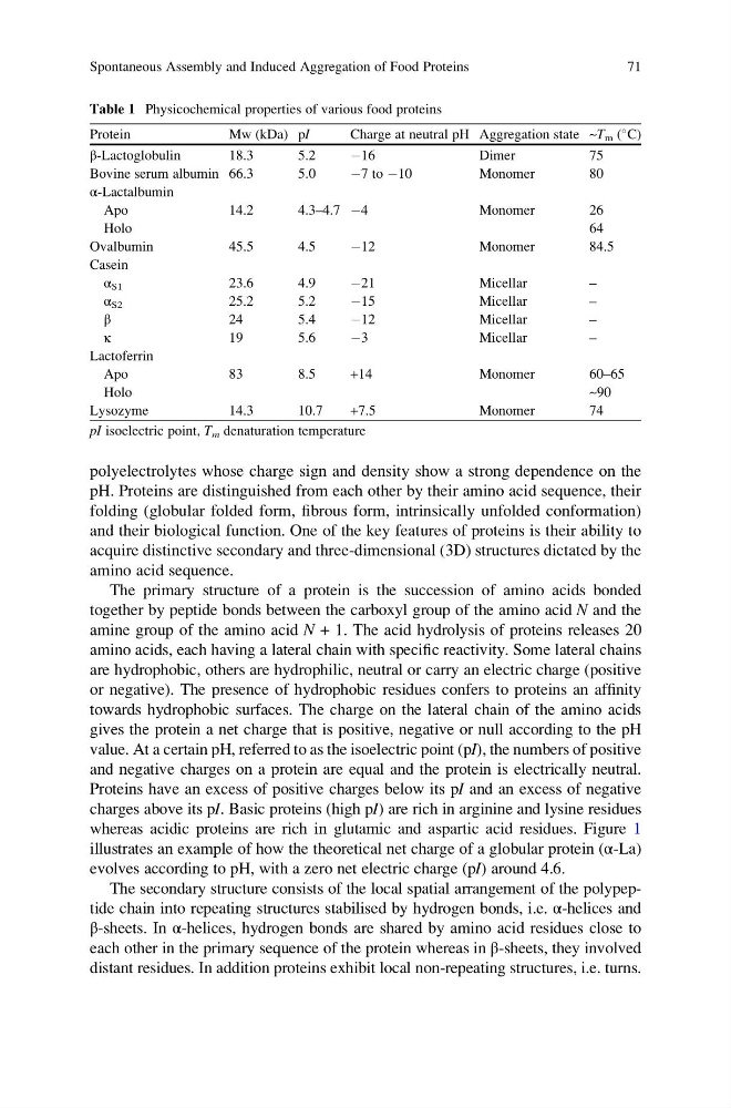
Table 1 Physicochemical properties of various food proteins
Protein
Mw (kDa) pI
Charge at neutral pH Aggregation state ~T m (
C)
β-Lactoglobulin
18.3
5.2
À16
Dimer
75
Bovine serum albumin 66.3
5.0
À7 to À10
Monomer
80
α-Lactalbumin
Apo
14.2
4.3–4.7 À4
Monomer
26
Holo
64
Ovalbumin
45.5
4.5
À12
Monomer
84.5
Casein
α S1
23.6
4.9
À21
Micellar
–
α S2
25.2
5.2
À15
Micellar
–
β
24
5.4
À12
Micellar
–
κ
19
5.6
À3
Micellar
–
Lactoferrin
Apo
83
8.5
+14
Monomer
60–65
Holo
~90
Lysozyme
14.3
10.7
+7.5
Monomer
74
pI isoelectric point, T m denaturation temperature
Spontaneous Assembly and Induced Aggregation of Food Proteins
71
pH. Proteins are distinguished from each other by their amino acid sequence, their
folding (globular folded form, fibrous form, intrinsically unfolded conformation)
and their biological function. One of the key features of proteins is their ability to
acquire distinctive secondary and three-dimensional (3D) structures dictated by the
amino acid sequence.
The primary structure of a protein is the succession of amino acids bonded
together by peptide bonds between the carboxyl group of the amino acid N and the
amine group of the amino acid N + 1. The acid hydrolysis of proteins releases 20
amino acids, each having a lateral chain with specific reactivity. Some lateral chains
are hydrophobic, others are hydrophilic, neutral or carry an electric charge (positive
or negative). The presence of hydrophobic residues confers to proteins an affinity
towards hydrophobic surfaces. The charge on the lateral chain of the amino acids
gives the protein a net charge that is positive, negative or null according to the pH
value. At a certain pH, referred to as the isoelectric point (pI), the numbers of positive
and negative charges on a protein are equal and the protein is electrically neutral.
Proteins have an excess of positive charges below its pI and an excess of negative
charges above its pI. Basic proteins (high pI) are rich in arginine and lysine residues
whereas acidic proteins are rich in glutamic and aspartic acid residues. Figure 1
illustrates an example of how the theoretical net charge of a globular protein (α-La)
evolves according to pH, with a zero net electric charge (pI) around 4.6.
The secondary structure consists of the local spatial arrangement of the polypeptide chain into repeating structures stabilised by hydrogen bonds, i.e. α-helices and
β-sheets. In α-helices, hydrogen bonds are shared by amino acid residues close to
each other in the primary sequence of the protein whereas in β-sheets, they involved
distant residues. In addition proteins exhibit local non-repeating structures, i.e. turns.
Table 1 Physicochemical properties of various food proteins
Protein
Mw (kDa) pI
Charge at neutral pH Aggregation state ~T m (
C)
β-Lactoglobulin
18.3
5.2
À16
Dimer
75
Bovine serum albumin 66.3
5.0
À7 to À10
Monomer
80
α-Lactalbumin
Apo
14.2
4.3–4.7 À4
Monomer
26
Holo
64
Ovalbumin
45.5
4.5
À12
Monomer
84.5
Casein
α S1
23.6
4.9
À21
Micellar
–
α S2
25.2
5.2
À15
Micellar
–
β
24
5.4
À12
Micellar
–
κ
19
5.6
À3
Micellar
–
Lactoferrin
Apo
83
8.5
+14
Monomer
60–65
Holo
~90
Lysozyme
14.3
10.7
+7.5
Monomer
74
pI isoelectric point, T m denaturation temperature
Spontaneous Assembly and Induced Aggregation of Food Proteins
71