

2.1.3 Transmission Electron Microscopy
The first low resolution TEM images of PEC phases can be found in the review
article of Philipp et al. [12] and a research article of Tiersch et al. [35]. Therein, TEM
images of cast pure carboxymethylcellulose (CMC) featuring fibrillar network
structures are compared with those of a CMC/PDADMAC PEC sample featuring
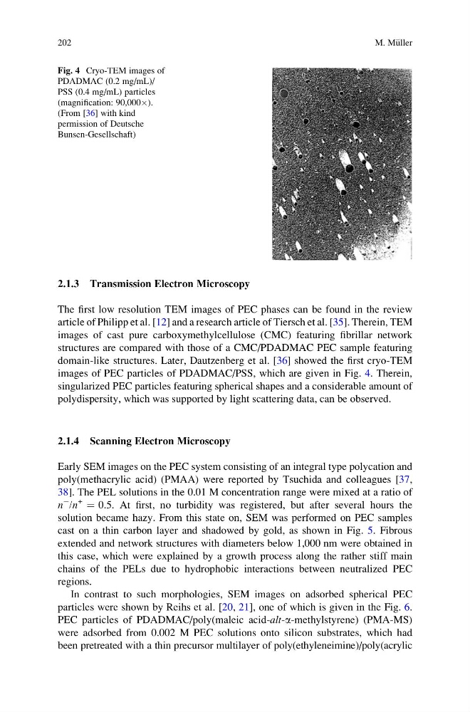
domain-like structures. Later, Dautzenberg et al. [36] showed the first cryo-TEM
images of PEC particles of PDADMAC/PSS, which are given in Fig. 4. Therein,
singularized PEC particles featuring spherical shapes and a considerable amount of
polydispersity, which was supported by light scattering data, can be observed.
2.1.4 Scanning Electron Microscopy
Early SEM images on the PEC system consisting of an integral type polycation and
poly(methacrylic acid) (PMAA) were reported by Tsuchida and colleagues [37,
38]. The PEL solutions in the 0.01 M concentration range were mixed at a ratio of
n
À /n
+
¼ 0.5. At first, no turbidity was registered, but after several hours the
solution became hazy. From this state on, SEM was performed on PEC samples
cast on a thin carbon layer and shadowed by gold, as shown in Fig. 5. Fibrous
extended and network structures with diameters below 1,000 nm were obtained in
this case, which were explained by a growth process along the rather stiff main
chains of the PELs due to hydrophobic interactions between neutralized PEC
regions.
In contrast to such morphologies, SEM images on adsorbed spherical PEC
particles were shown by Reihs et al. [20, 21], one of which is given in the Fig. 6.
PEC particles of PDADMAC/poly(maleic acid-alt-a-methylstyrene) (PMA-MS)
were adsorbed from 0.002 M PEC solutions onto silicon substrates, which had
been pretreated with a thin precursor multilayer of poly(ethyleneimine)/poly(acrylic
Fig. 4 Cryo-TEM images of
PDADMAC (0.2 mg/mL)/
PSS (0.4 mg/mL) particles
(magnification: 90,000Â).
(From [36] with kind
permission of Deutsche
Bunsen-Gesellschaft)
202
M. M€ uller
The first low resolution TEM images of PEC phases can be found in the review
article of Philipp et al. [12] and a research article of Tiersch et al. [35]. Therein, TEM
images of cast pure carboxymethylcellulose (CMC) featuring fibrillar network
structures are compared with those of a CMC/PDADMAC PEC sample featuring
domain-like structures. Later, Dautzenberg et al. [36] showed the first cryo-TEM
images of PEC particles of PDADMAC/PSS, which are given in Fig. 4. Therein,
singularized PEC particles featuring spherical shapes and a considerable amount of
polydispersity, which was supported by light scattering data, can be observed.
2.1.4 Scanning Electron Microscopy
Early SEM images on the PEC system consisting of an integral type polycation and
poly(methacrylic acid) (PMAA) were reported by Tsuchida and colleagues [37,
38]. The PEL solutions in the 0.01 M concentration range were mixed at a ratio of
n
À /n
+
¼ 0.5. At first, no turbidity was registered, but after several hours the
solution became hazy. From this state on, SEM was performed on PEC samples
cast on a thin carbon layer and shadowed by gold, as shown in Fig. 5. Fibrous
extended and network structures with diameters below 1,000 nm were obtained in
this case, which were explained by a growth process along the rather stiff main
chains of the PELs due to hydrophobic interactions between neutralized PEC
regions.
In contrast to such morphologies, SEM images on adsorbed spherical PEC
particles were shown by Reihs et al. [20, 21], one of which is given in the Fig. 6.
PEC particles of PDADMAC/poly(maleic acid-alt-a-methylstyrene) (PMA-MS)
were adsorbed from 0.002 M PEC solutions onto silicon substrates, which had
been pretreated with a thin precursor multilayer of poly(ethyleneimine)/poly(acrylic
Fig. 4 Cryo-TEM images of
PDADMAC (0.2 mg/mL)/
PSS (0.4 mg/mL) particles
(magnification: 90,000Â).
(From [36] with kind
permission of Deutsche
Bunsen-Gesellschaft)
202
M. M€ uller