

include Young’s modulus and stress relaxation properties. These parameters have
been discussed in the section on mechanical properties (Sect. 3.1).
The aortic heart valve, which controls the flow of oxygenated blood from the left
ventricle into the ascending aorta for distribution to the whole body, is prone to
failure. The most common types of artificial replacement are the mechanical and
the bioprosthetic heart valves, but they both have shortcomings. One of the early
attempts to use PVA-C to alleviate the mechanical problems that can lead to tissue
tearing, calcification, and eventual failure of the bioprosthetic heart valve addressed
the lack of expansibility of the mounting stent [7]. The PVA solution composition
and freeze–thaw cycling processing conditions for the preparation of PVA-C that
best mimics the mechanical properties of the porcine aortic root were determined. A
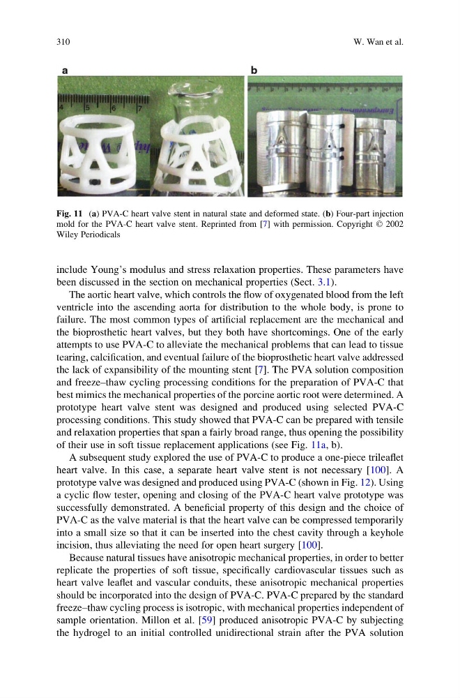
prototype heart valve stent was designed and produced using selected PVA-C
processing conditions. This study showed that PVA-C can be prepared with tensile
and relaxation properties that span a fairly broad range, thus opening the possibility
of their use in soft tissue replacement applications (see Fig. 11a, b).
A subsequent study explored the use of PVA-C to produce a one-piece trileaflet
heart valve. In this case, a separate heart valve stent is not necessary [100]. A
prototype valve was designed and produced using PVA-C (shown in Fig. 12). Using
a cyclic flow tester, opening and closing of the PVA-C heart valve prototype was
successfully demonstrated. A beneficial property of this design and the choice of
PVA-C as the valve material is that the heart valve can be compressed temporarily
into a small size so that it can be inserted into the chest cavity through a keyhole
incision, thus alleviating the need for open heart surgery [100].
Because natural tissues have anisotropic mechanical properties, in order to better
replicate the properties of soft tissue, specifically cardiovascular tissues such as
heart valve leaflet and vascular conduits, these anisotropic mechanical properties
should be incorporated into the design of PVA-C. PVA-C prepared by the standard
freeze–thaw cycling process is isotropic, with mechanical properties independent of
sample orientation. Millon et al. [59] produced anisotropic PVA-C by subjecting
the hydrogel to an initial controlled unidirectional strain after the PVA solution
Fig. 11 (a) PVA-C heart valve stent in natural state and deformed state. (b) Four-part injection
mold for the PVA-C heart valve stent. Reprinted from [7] with permission. Copyright © 2002
Wiley Periodicals
310
W. Wan et al.
been discussed in the section on mechanical properties (Sect. 3.1).
The aortic heart valve, which controls the flow of oxygenated blood from the left
ventricle into the ascending aorta for distribution to the whole body, is prone to
failure. The most common types of artificial replacement are the mechanical and
the bioprosthetic heart valves, but they both have shortcomings. One of the early
attempts to use PVA-C to alleviate the mechanical problems that can lead to tissue
tearing, calcification, and eventual failure of the bioprosthetic heart valve addressed
the lack of expansibility of the mounting stent [7]. The PVA solution composition
and freeze–thaw cycling processing conditions for the preparation of PVA-C that
best mimics the mechanical properties of the porcine aortic root were determined. A
prototype heart valve stent was designed and produced using selected PVA-C
processing conditions. This study showed that PVA-C can be prepared with tensile
and relaxation properties that span a fairly broad range, thus opening the possibility
of their use in soft tissue replacement applications (see Fig. 11a, b).
A subsequent study explored the use of PVA-C to produce a one-piece trileaflet
heart valve. In this case, a separate heart valve stent is not necessary [100]. A
prototype valve was designed and produced using PVA-C (shown in Fig. 12). Using
a cyclic flow tester, opening and closing of the PVA-C heart valve prototype was
successfully demonstrated. A beneficial property of this design and the choice of
PVA-C as the valve material is that the heart valve can be compressed temporarily
into a small size so that it can be inserted into the chest cavity through a keyhole
incision, thus alleviating the need for open heart surgery [100].
Because natural tissues have anisotropic mechanical properties, in order to better
replicate the properties of soft tissue, specifically cardiovascular tissues such as
heart valve leaflet and vascular conduits, these anisotropic mechanical properties
should be incorporated into the design of PVA-C. PVA-C prepared by the standard
freeze–thaw cycling process is isotropic, with mechanical properties independent of
sample orientation. Millon et al. [59] produced anisotropic PVA-C by subjecting
the hydrogel to an initial controlled unidirectional strain after the PVA solution
Fig. 11 (a) PVA-C heart valve stent in natural state and deformed state. (b) Four-part injection
mold for the PVA-C heart valve stent. Reprinted from [7] with permission. Copyright © 2002
Wiley Periodicals
310
W. Wan et al.