

When using cryogels, a much more open structure is used, which reduces the risk
that cells will be bound from different angles. However, in cryogels also, there is a
risk for too-strong binding. One mode of operation would be to operate with very low
substitution on the gel matrix. This is a sensitive issue since a too low a degree of
derivatization would lead to no capturing of cells at all. Since the firmness of binding
to some extent is a function of time, it is possible to modulate the time of contact and
thereby facilitate release of bound cells. Another mode of operation is to work under
conditions where binding is weakened, e.g., by modulating the pH or by adding a
competing ligand in free solution that may interact with the cell surface, thereby
blocking, or at least reducing, the possibilities for optimal binding to the support.
Cells have been separated on cryogels. The initial studies were carried out on
bacterial cells because they are robust and easier to handle than mammalian cells. In
this context, it needs to be stressed that the diffusion rate of cells is extremely low.
In order to facilitate binding of cells to the adsorbent, the flow through the column
was stopped for a short time before continuing the buffer flow.
In initial studies to demonstrate that it is possible to capture cells and then
successfully harvest them, an anion-exchanger was used [54]. The monomers
used for producing the cryogel were acrylamide, N,N
0 -methylenebisacrylamide,
and the charged monomer 2-(dimethylamino)ethyl methacrylate. The concentration
of charged groups in the final cryogel was approximately 1,000 μmol of tertiary
amino groups per gram of dry gel. A gel matrix of 5 mL constituted one column,
and initially a trial was made to verify that microbial cells could pass through the
gel. The eluting medium was HEPES buffer, 20 mM pH 7.0 containing1 M NaCl.
The high concentration of salt was sufficient to suppress electrostatic interactions.
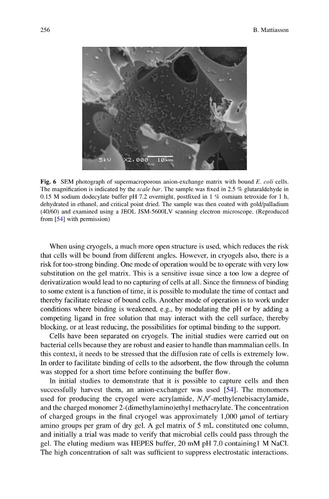
Fig. 6 SEM photograph of supermacroporous anion-exchange matrix with bound E. coli cells.
The magnification is indicated by the scale bar. The sample was fixed in 2.5 % glutaraldehyde in
0.15 M sodium dodecylate buffer pH 7.2 overnight, postfixed in 1 % osmium tetroxide for 1 h,
dehydrated in ethanol, and critical point dried. The sample was then coated with gold/palladium
(40/60) and examined using a JEOL JSM-5600LV scanning electron microscope. (Reproduced
from [54] with permission)
256
B. Mattiasson
that cells will be bound from different angles. However, in cryogels also, there is a
risk for too-strong binding. One mode of operation would be to operate with very low
substitution on the gel matrix. This is a sensitive issue since a too low a degree of
derivatization would lead to no capturing of cells at all. Since the firmness of binding
to some extent is a function of time, it is possible to modulate the time of contact and
thereby facilitate release of bound cells. Another mode of operation is to work under
conditions where binding is weakened, e.g., by modulating the pH or by adding a
competing ligand in free solution that may interact with the cell surface, thereby
blocking, or at least reducing, the possibilities for optimal binding to the support.
Cells have been separated on cryogels. The initial studies were carried out on
bacterial cells because they are robust and easier to handle than mammalian cells. In
this context, it needs to be stressed that the diffusion rate of cells is extremely low.
In order to facilitate binding of cells to the adsorbent, the flow through the column
was stopped for a short time before continuing the buffer flow.
In initial studies to demonstrate that it is possible to capture cells and then
successfully harvest them, an anion-exchanger was used [54]. The monomers
used for producing the cryogel were acrylamide, N,N
0 -methylenebisacrylamide,
and the charged monomer 2-(dimethylamino)ethyl methacrylate. The concentration
of charged groups in the final cryogel was approximately 1,000 μmol of tertiary
amino groups per gram of dry gel. A gel matrix of 5 mL constituted one column,
and initially a trial was made to verify that microbial cells could pass through the
gel. The eluting medium was HEPES buffer, 20 mM pH 7.0 containing1 M NaCl.
The high concentration of salt was sufficient to suppress electrostatic interactions.
Fig. 6 SEM photograph of supermacroporous anion-exchange matrix with bound E. coli cells.
The magnification is indicated by the scale bar. The sample was fixed in 2.5 % glutaraldehyde in
0.15 M sodium dodecylate buffer pH 7.2 overnight, postfixed in 1 % osmium tetroxide for 1 h,
dehydrated in ethanol, and critical point dried. The sample was then coated with gold/palladium
(40/60) and examined using a JEOL JSM-5600LV scanning electron microscope. (Reproduced
from [54] with permission)
256
B. Mattiasson