

ammonium ion. The results were encouraging, with a high productivity of monoclonal antibodies, almost equal to that of the best optimized medium. Most studies
on culturing of mammalian cells are devoted towards tissue engineering, which is
outside the scope of this review.
7 Chromatographic Separation of Particulate Matter
7.1 Cells
Compared to separation of soluble biomolecules, cell separation presents some
additional challenges. Cells have surfaces where many points of possible interaction with an affinity matrix are present. This means that when a cell comes into
contact with a solid support, several interactions may take place simultaneously, or
within a very short time frame. That, in turn, leads to a much firmer binding than
can be expected on the basis of the strength of the individual binding. The result of
this is that cells get stuck on the column and are very difficult to elute out in viable
shape. This has been the situation when columns packed with chromatographic
beds have been used with the intention of separating cells.
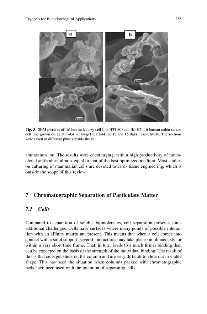
Fig. 5 SEM pictures of (a) human kidney cell line HT1080 and (b) HT116 human colon cancer
cell line grown on gelatin-AAm cryogel scaffold for 18 and 15 days, respectively. The sections
were taken at different places inside the gel
Cryogels for Biotechnological Applications
255
on culturing of mammalian cells are devoted towards tissue engineering, which is
outside the scope of this review.
7 Chromatographic Separation of Particulate Matter
7.1 Cells
Compared to separation of soluble biomolecules, cell separation presents some
additional challenges. Cells have surfaces where many points of possible interaction with an affinity matrix are present. This means that when a cell comes into
contact with a solid support, several interactions may take place simultaneously, or
within a very short time frame. That, in turn, leads to a much firmer binding than
can be expected on the basis of the strength of the individual binding. The result of
this is that cells get stuck on the column and are very difficult to elute out in viable
shape. This has been the situation when columns packed with chromatographic
beds have been used with the intention of separating cells.
Fig. 5 SEM pictures of (a) human kidney cell line HT1080 and (b) HT116 human colon cancer
cell line grown on gelatin-AAm cryogel scaffold for 18 and 15 days, respectively. The sections
were taken at different places inside the gel
Cryogels for Biotechnological Applications
255