

microphase jellifies, preventing attainment of thermodynamic equilibrium
[77]. The formation of the gel microphase may be determined by aggregation of
PVA chains in a confined environment, which is driven by hydrogen bonding,
consequent formation of crystallites, and vitrification of the swollen amorphous
phase at a temperature that is probably below the glass transition [4–6]. This
scenario holds both for solutions with PVA volume fraction Φ ! 0.086 and for
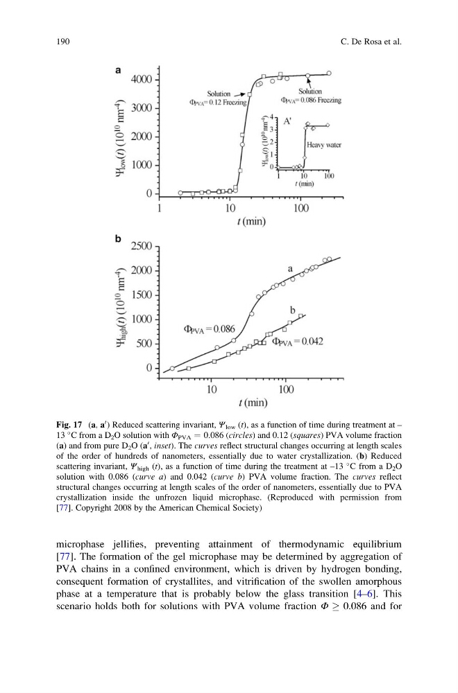
Fig. 17 (a, a
0 ) Reduced scattering invariant, Ψ low (t), as a function of time during treatment at –
13
C from a D 2 O solution with Φ PVA ¼ 0.086 (circles) and 0.12 (squares) PVA volume fraction
(a) and from pure D 2 O (a
0 , inset). The curves reflect structural changes occurring at length scales
of the order of hundreds of nanometers, essentially due to water crystallization. (b) Reduced
scattering invariant, Ψ high (t), as a function of time during the treatment at –13
C from a D 2 O
solution with 0.086 (curve a) and 0.042 (curve b) PVA volume fraction. The curves reflect
structural changes occurring at length scales of the order of nanometers, essentially due to PVA
crystallization inside the unfrozen liquid microphase. (Reproduced with permission from
[77]. Copyright 2008 by the American Chemical Society)
190
C. De Rosa et al.
[77]. The formation of the gel microphase may be determined by aggregation of
PVA chains in a confined environment, which is driven by hydrogen bonding,
consequent formation of crystallites, and vitrification of the swollen amorphous
phase at a temperature that is probably below the glass transition [4–6]. This
scenario holds both for solutions with PVA volume fraction Φ ! 0.086 and for
Fig. 17 (a, a
0 ) Reduced scattering invariant, Ψ low (t), as a function of time during treatment at –
13
C from a D 2 O solution with Φ PVA ¼ 0.086 (circles) and 0.12 (squares) PVA volume fraction
(a) and from pure D 2 O (a
0 , inset). The curves reflect structural changes occurring at length scales
of the order of hundreds of nanometers, essentially due to water crystallization. (b) Reduced
scattering invariant, Ψ high (t), as a function of time during the treatment at –13
C from a D 2 O
solution with 0.086 (curve a) and 0.042 (curve b) PVA volume fraction. The curves reflect
structural changes occurring at length scales of the order of nanometers, essentially due to PVA
crystallization inside the unfrozen liquid microphase. (Reproduced with permission from
[77]. Copyright 2008 by the American Chemical Society)
190
C. De Rosa et al.