

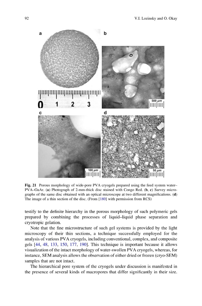
testify to the definite hierarchy in the porous morphology of such polymeric gels
prepared by combining the processes of liquid–liquid phase separation and
cryotropic gelation.
Note that the fine microstructure of such gel systems is provided by the light
microscopy of their thin sections, a technique successfully employed for the
analysis of various PVA cryogels, including conventional, complex, and composite
gels [44, 48, 133, 150, 177, 190]. This technique is important because it allows
visualization of the intact morphology of water-swollen PVA cryogels, whereas, for
instance, SEM analysis allows the observation of either dried or frozen (cryo-SEM)
samples that are not intact.
The hierarchical pore system of the cryogels under discussion is manifested in
the presence of several kinds of macropores that differ significantly in their size.
Fig. 21 Porous morphology of wide-pore PVA cryogels prepared using the feed system water–
PVA–GuAr. (a) Photograph of 2-mm-thick disc stained with Congo Red. (b, c) Survey micrographs of the same disc obtained with an optical microscope at two different magnifications. (d)
The image of a thin section of the disc. (From [180] with permission from RCS)
92
V.I. Lozinsky and O. Okay
prepared by combining the processes of liquid–liquid phase separation and
cryotropic gelation.
Note that the fine microstructure of such gel systems is provided by the light
microscopy of their thin sections, a technique successfully employed for the
analysis of various PVA cryogels, including conventional, complex, and composite
gels [44, 48, 133, 150, 177, 190]. This technique is important because it allows
visualization of the intact morphology of water-swollen PVA cryogels, whereas, for
instance, SEM analysis allows the observation of either dried or frozen (cryo-SEM)
samples that are not intact.
The hierarchical pore system of the cryogels under discussion is manifested in
the presence of several kinds of macropores that differ significantly in their size.
Fig. 21 Porous morphology of wide-pore PVA cryogels prepared using the feed system water–
PVA–GuAr. (a) Photograph of 2-mm-thick disc stained with Congo Red. (b, c) Survey micrographs of the same disc obtained with an optical microscope at two different magnifications. (d)
The image of a thin section of the disc. (From [180] with permission from RCS)
92
V.I. Lozinsky and O. Okay