

transmission electron microscopy (TEM), (Sabo-Attwood
et al. 2012) showed that 3.5-nm AuNPs entered Nicotiana
xanthi through the roots and moved into the vascular system.
Aggregates of 18-nm AuNPs were detected only in the root
cell cytoplasm. Exposure to small particles led to leaf
necrosis after 14 days, but with large particles, no differences from the control were observed. At very high AuNP
concentrations, no physiological effect has been described in
Glycine max L. (Falco et al. 2011) and aquatic aquarium
plants (Glenn et al. 2012).
Several studies on the mechanisms of nanoparticle entry
into plants and on nanoparticle phytotoxicity (Taylor 2011)
have been done with Arabidopsis thaliana, a classical object
in current plant physiology. The addition of 24-nm AuNPs
(10–80 mg l
−1 ) to the growth medium led to a threefold
increase in total seed yield, as compared with the control. It
also markedly increased the length and diameter of the stem
and roots. Exciting results came from the study by Taylor
et al. (2014), who found that the root length in A. thaliana
grown on an agar medium with 100 mg l
−1 of K(AuCl4)
was reduced by 75%. But there also was a slight decrease in
the expression of genes coding for aquaporins and proteins
implicated in the transport metal ions such as copper,
cadmium, iron, and nickel ions. Oxidized gold was found
simultaneously in the vascular system of roots and shoots of
plant species A. thaliana, but AuNPs synthesized in plants
were detected only in root tissues. Gold chlorides were much
more genotoxic than AuNPs (Taylor et al. 2014). Overall,
the toxicity of metal ions was much higher than that of
nanoparticles, and AgNPs were more phytotoxic than
AuNPs (Notter et al. 2014).
In a study of the toxicity of metallic nanoparticles to
callus cultures, Fazal et al. (2016) showed that AuNPs
enhance callus proliferation in Prunella vulgaris (L.). Cellular entry and toxicity of nanoparticles are often investigated with suspension cultures (Alkilany et al. 2010).
Suspension cultures of plant cells are more sensitive to a
broad range of compounds and abiotic effects (Rains et al.
1989). Biochemical and physiological responses develop
within a short time and are fairly evenly distributed across
the population, unlike what is observed in a whole plant or
its organs. Additionally, one can expect that the effects of
nanoparticles on suspension culture cells will be more significant owing to the absence of specialized protective
structures such as cuticles or epidermis. The addition of
20-nm AuNPs to the growth medium of A. thaliana
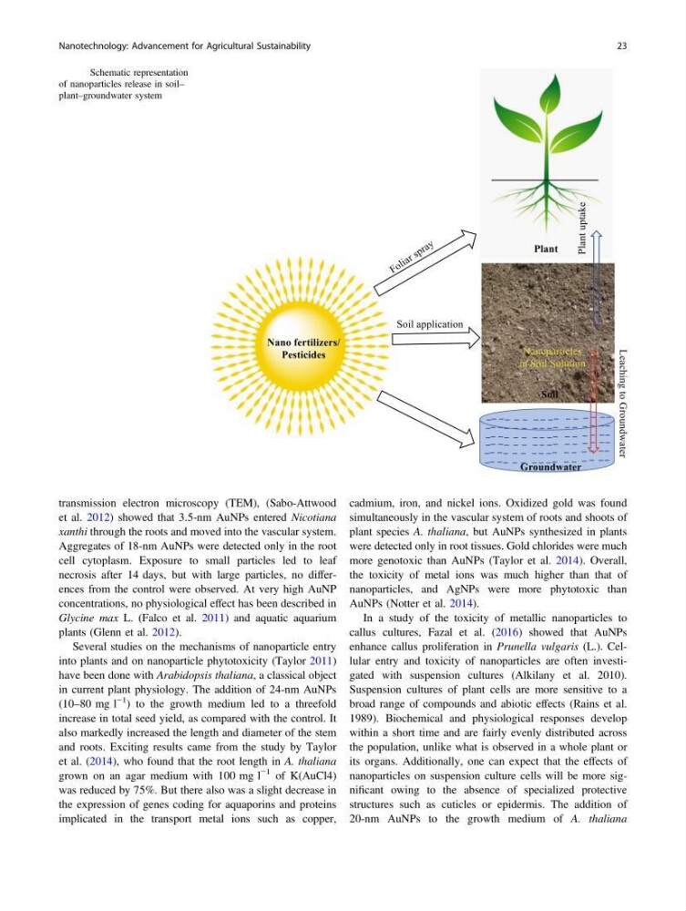
Fig. 2 Schematic representation
of nanoparticles release in soil–
plant–groundwater system
Nanotechnology: Advancement for Agricultural Sustainability
23
et al. 2012) showed that 3.5-nm AuNPs entered Nicotiana
xanthi through the roots and moved into the vascular system.
Aggregates of 18-nm AuNPs were detected only in the root
cell cytoplasm. Exposure to small particles led to leaf
necrosis after 14 days, but with large particles, no differences from the control were observed. At very high AuNP
concentrations, no physiological effect has been described in
Glycine max L. (Falco et al. 2011) and aquatic aquarium
plants (Glenn et al. 2012).
Several studies on the mechanisms of nanoparticle entry
into plants and on nanoparticle phytotoxicity (Taylor 2011)
have been done with Arabidopsis thaliana, a classical object
in current plant physiology. The addition of 24-nm AuNPs
(10–80 mg l
−1 ) to the growth medium led to a threefold
increase in total seed yield, as compared with the control. It
also markedly increased the length and diameter of the stem
and roots. Exciting results came from the study by Taylor
et al. (2014), who found that the root length in A. thaliana
grown on an agar medium with 100 mg l
−1 of K(AuCl4)
was reduced by 75%. But there also was a slight decrease in
the expression of genes coding for aquaporins and proteins
implicated in the transport metal ions such as copper,
cadmium, iron, and nickel ions. Oxidized gold was found
simultaneously in the vascular system of roots and shoots of
plant species A. thaliana, but AuNPs synthesized in plants
were detected only in root tissues. Gold chlorides were much
more genotoxic than AuNPs (Taylor et al. 2014). Overall,
the toxicity of metal ions was much higher than that of
nanoparticles, and AgNPs were more phytotoxic than
AuNPs (Notter et al. 2014).
In a study of the toxicity of metallic nanoparticles to
callus cultures, Fazal et al. (2016) showed that AuNPs
enhance callus proliferation in Prunella vulgaris (L.). Cellular entry and toxicity of nanoparticles are often investigated with suspension cultures (Alkilany et al. 2010).
Suspension cultures of plant cells are more sensitive to a
broad range of compounds and abiotic effects (Rains et al.
1989). Biochemical and physiological responses develop
within a short time and are fairly evenly distributed across
the population, unlike what is observed in a whole plant or
its organs. Additionally, one can expect that the effects of
nanoparticles on suspension culture cells will be more significant owing to the absence of specialized protective
structures such as cuticles or epidermis. The addition of
20-nm AuNPs to the growth medium of A. thaliana
Fig. 2 Schematic representation
of nanoparticles release in soil–
plant–groundwater system
Nanotechnology: Advancement for Agricultural Sustainability
23