

488
A. Chatterjee et al.
0
10
20
30
40
50
60
70
80
340 350 360 370 380 390 400 410 420 429 430 440
OD Value
Wavelength(nm)
Peak of synthesized silver nanoparƟcles from Haldi
Peak of silver nanoparƟcles from Neem
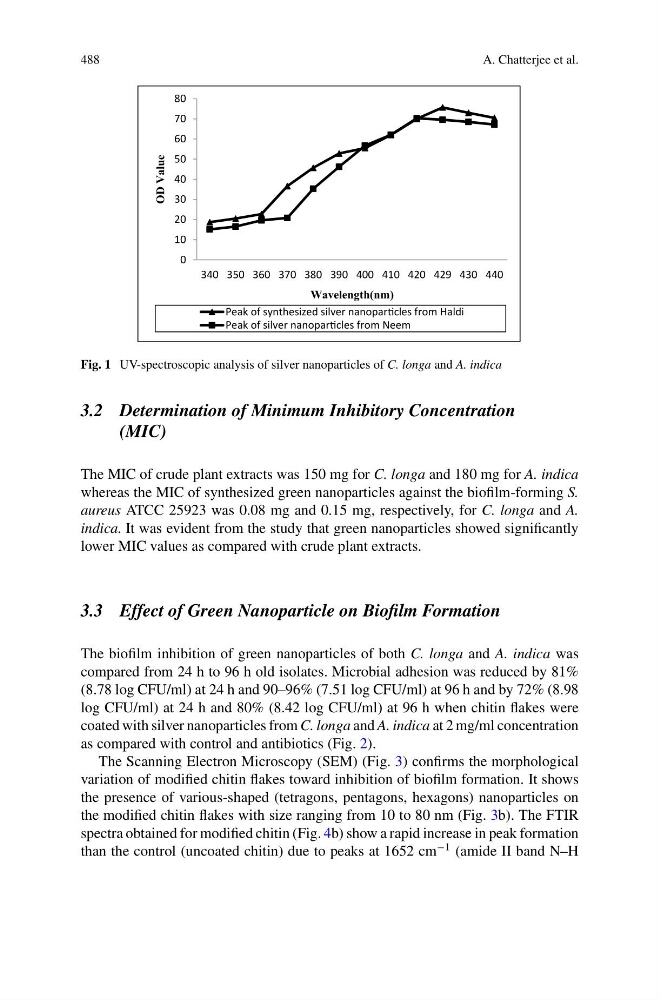
Fig. 1 UV-spectroscopic analysis of silver nanoparticles of C. longa and A. indica
3.2 Determination of Minimum Inhibitory Concentration
(MIC)
The MIC of crude plant extracts was 150 mg for C. longa and 180 mg for A. indica
whereas the MIC of synthesized green nanoparticles against the biofilm-forming S.
aureus ATCC 25923 was 0.08 mg and 0.15 mg, respectively, for C. longa and A.
indica. It was evident from the study that green nanoparticles showed significantly
lower MIC values as compared with crude plant extracts.
3.3 Effect of Green Nanoparticle on Biofilm Formation
The biofilm inhibition of green nanoparticles of both C. longa and A. indica was
compared from 24 h to 96 h old isolates. Microbial adhesion was reduced by 81%
(8.78 log CFU/ml) at 24 h and 90–96% (7.51 log CFU/ml) at 96 h and by 72% (8.98
log CFU/ml) at 24 h and 80% (8.42 log CFU/ml) at 96 h when chitin flakes were
coated with silver nanoparticles from C. longa and A. indica at 2 mg/ml concentration
as compared with control and antibiotics (Fig. 2).
The Scanning Electron Microscopy (SEM) (Fig. 3) confirms the morphological
variation of modified chitin flakes toward inhibition of biofilm formation. It shows
the presence of various-shaped (tetragons, pentagons, hexagons) nanoparticles on
the modified chitin flakes with size ranging from 10 to 80 nm (Fig. 3b). The FTIR
spectra obtained for modified chitin (Fig. 4b) show a rapid increase in peak formation
than the control (uncoated chitin) due to peaks at 1652 cm
−1 (amide II band N–H
A. Chatterjee et al.
0
10
20
30
40
50
60
70
80
340 350 360 370 380 390 400 410 420 429 430 440
OD Value
Wavelength(nm)
Peak of synthesized silver nanoparƟcles from Haldi
Peak of silver nanoparƟcles from Neem
Fig. 1 UV-spectroscopic analysis of silver nanoparticles of C. longa and A. indica
3.2 Determination of Minimum Inhibitory Concentration
(MIC)
The MIC of crude plant extracts was 150 mg for C. longa and 180 mg for A. indica
whereas the MIC of synthesized green nanoparticles against the biofilm-forming S.
aureus ATCC 25923 was 0.08 mg and 0.15 mg, respectively, for C. longa and A.
indica. It was evident from the study that green nanoparticles showed significantly
lower MIC values as compared with crude plant extracts.
3.3 Effect of Green Nanoparticle on Biofilm Formation
The biofilm inhibition of green nanoparticles of both C. longa and A. indica was
compared from 24 h to 96 h old isolates. Microbial adhesion was reduced by 81%
(8.78 log CFU/ml) at 24 h and 90–96% (7.51 log CFU/ml) at 96 h and by 72% (8.98
log CFU/ml) at 24 h and 80% (8.42 log CFU/ml) at 96 h when chitin flakes were
coated with silver nanoparticles from C. longa and A. indica at 2 mg/ml concentration
as compared with control and antibiotics (Fig. 2).
The Scanning Electron Microscopy (SEM) (Fig. 3) confirms the morphological
variation of modified chitin flakes toward inhibition of biofilm formation. It shows
the presence of various-shaped (tetragons, pentagons, hexagons) nanoparticles on
the modified chitin flakes with size ranging from 10 to 80 nm (Fig. 3b). The FTIR
spectra obtained for modified chitin (Fig. 4b) show a rapid increase in peak formation
than the control (uncoated chitin) due to peaks at 1652 cm
−1 (amide II band N–H