

100
S. E. H. Murph and A. Goriounova
nanoparticles prepared in the absence of stainless-steel wool (Fig. 3a) or presence of
stainless-steel wool, namely 25 ± 5 nm average (Fig. 3b) gold nanoparticles.
These differences in sizes could be attributed to the unique stainless-steel wool
topography. When investigated under a scanning electron microscope, one could see
that the stainless-steel wool control has distinct valleys, steps, and crevices (Fig. 3gi). The presence of nano-features on stainless-steel wool surfaces plays a crucial role
in the nucleation and growth of metallic nanoparticles.
Typically, gold nanoparticles’ nucleation and growth mechanism are based on
the following steps: (1) reduction of the gold precursor and formation of nucleation
centers clusters, (2) formation of seed particles, and (3) slow and fast growth of gold
nanoparticles [1, 4, 5]. The support’s surface condition, topography, and roughness
generate different gold nanoparticle morphologies and surface loading, e.g. surface
densities. A highly defective surface facilitates clusters’ nucleation and growth at
point defects on the terraces [1, 16]. The stainless-steel wool grooves have a diameter range of 8–45 nm with an average of 26 nm. We suggest that the grooves and
roughness of the stainless-steel wool surface blocked and hindered the growth of
larger gold nanoparticles (Fig. 3g-i).
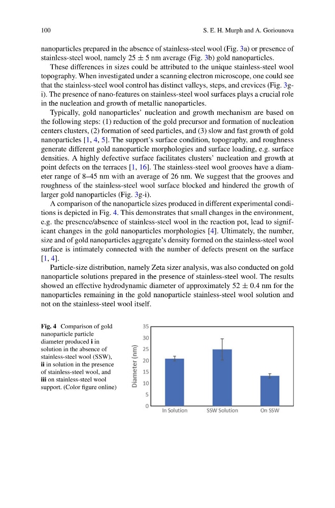
A comparison of the nanoparticle sizes produced in different experimental conditions is depicted in Fig. 4. This demonstrates that small changes in the environment,
e.g. the presence/absence of stainless-steel wool in the reaction pot, lead to significant changes in the gold nanoparticles morphologies [4]. Ultimately, the number,
size and of gold nanoparticles aggregate’s density formed on the stainless-steel wool
surface is intimately connected with the number of defects present on the surface
[1, 4].
Particle-size distribution, namely Zeta sizer analysis, was also conducted on gold
nanoparticle solutions prepared in the presence of stainless-steel wool. The results
showed an effective hydrodynamic diameter of approximately 52 ± 0.4 nm for the
nanoparticles remaining in the gold nanoparticle stainless-steel wool solution and
not on the stainless-steel wool itself.
Fig. 4 Comparison of gold
nanoparticle particle
diameter produced i in
solution in the absence of
stainless-steel wool (SSW),
ii in solution in the presence
of stainless-steel wool, and
iii on stainless-steel wool
support. (Color figure online)
S. E. H. Murph and A. Goriounova
nanoparticles prepared in the absence of stainless-steel wool (Fig. 3a) or presence of
stainless-steel wool, namely 25 ± 5 nm average (Fig. 3b) gold nanoparticles.
These differences in sizes could be attributed to the unique stainless-steel wool
topography. When investigated under a scanning electron microscope, one could see
that the stainless-steel wool control has distinct valleys, steps, and crevices (Fig. 3gi). The presence of nano-features on stainless-steel wool surfaces plays a crucial role
in the nucleation and growth of metallic nanoparticles.
Typically, gold nanoparticles’ nucleation and growth mechanism are based on
the following steps: (1) reduction of the gold precursor and formation of nucleation
centers clusters, (2) formation of seed particles, and (3) slow and fast growth of gold
nanoparticles [1, 4, 5]. The support’s surface condition, topography, and roughness
generate different gold nanoparticle morphologies and surface loading, e.g. surface
densities. A highly defective surface facilitates clusters’ nucleation and growth at
point defects on the terraces [1, 16]. The stainless-steel wool grooves have a diameter range of 8–45 nm with an average of 26 nm. We suggest that the grooves and
roughness of the stainless-steel wool surface blocked and hindered the growth of
larger gold nanoparticles (Fig. 3g-i).
A comparison of the nanoparticle sizes produced in different experimental conditions is depicted in Fig. 4. This demonstrates that small changes in the environment,
e.g. the presence/absence of stainless-steel wool in the reaction pot, lead to significant changes in the gold nanoparticles morphologies [4]. Ultimately, the number,
size and of gold nanoparticles aggregate’s density formed on the stainless-steel wool
surface is intimately connected with the number of defects present on the surface
[1, 4].
Particle-size distribution, namely Zeta sizer analysis, was also conducted on gold
nanoparticle solutions prepared in the presence of stainless-steel wool. The results
showed an effective hydrodynamic diameter of approximately 52 ± 0.4 nm for the
nanoparticles remaining in the gold nanoparticle stainless-steel wool solution and
not on the stainless-steel wool itself.
Fig. 4 Comparison of gold
nanoparticle particle
diameter produced i in
solution in the absence of
stainless-steel wool (SSW),
ii in solution in the presence
of stainless-steel wool, and
iii on stainless-steel wool
support. (Color figure online)