

5.3 Search for Different Chemical Structure Types
165
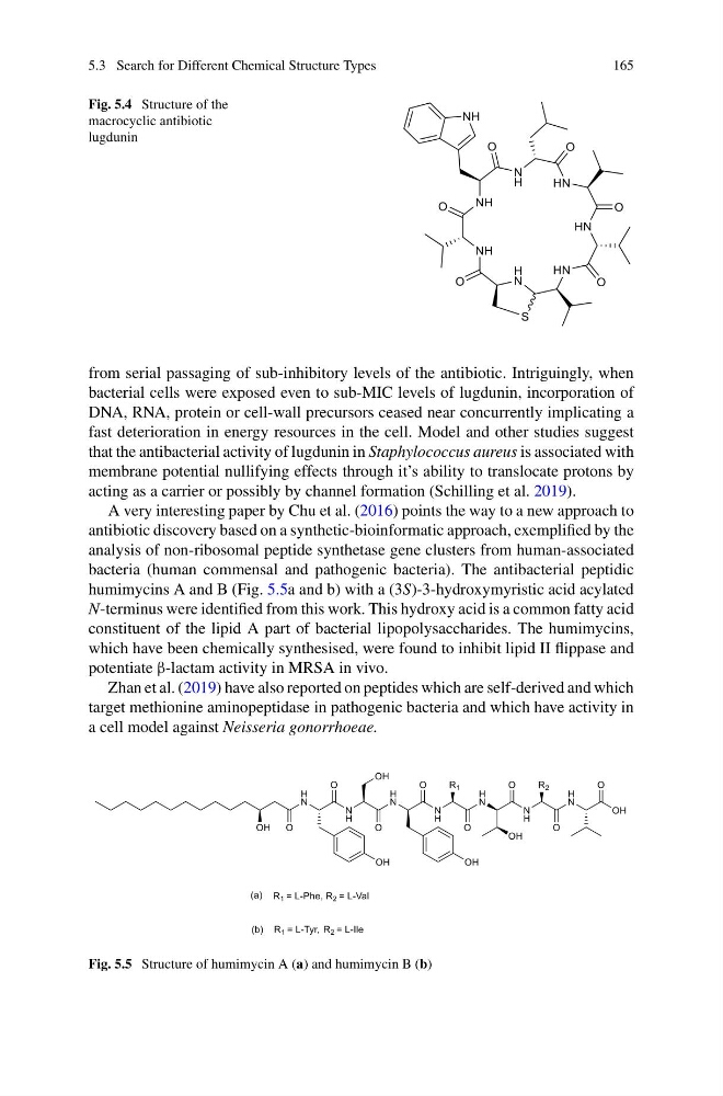
Fig. 5.4 Structure of the
macrocyclic antibiotic
lugdunin
from serial passaging of sub-inhibitory levels of the antibiotic. Intriguingly, when
bacterial cells were exposed even to sub-MIC levels of lugdunin, incorporation of
DNA, RNA, protein or cell-wall precursors ceased near concurrently implicating a
fast deterioration in energy resources in the cell. Model and other studies suggest
that the antibacterial activity of lugdunin in Staphylococcus aureus is associated with
membrane potential nullifying effects through it’s ability to translocate protons by
acting as a carrier or possibly by channel formation (Schilling et al. 2019).
A very interesting paper by Chu et al. (2016) points the way to a new approach to
antibiotic discovery based on a synthetic-bioinformatic approach, exemplified by the
analysis of non-ribosomal peptide synthetase gene clusters from human-associated
bacteria (human commensal and pathogenic bacteria). The antibacterial peptidic
humimycins A and B (Fig. 5.5a and b) with a (3S)-3-hydroxymyristic acid acylated
N-terminus were identified from this work. This hydroxy acid is a common fatty acid
constituent of the lipid A part of bacterial lipopolysaccharides. The humimycins,
which have been chemically synthesised, were found to inhibit lipid II flippase and
potentiate β-lactam activity in MRSA in vivo.
Zhan et al. (2019) have also reported on peptides which are self-derived and which
target methionine aminopeptidase in pathogenic bacteria and which have activity in
a cell model against Neisseria gonorrhoeae.
Fig. 5.5 Structure of humimycin A (a) and humimycin B (b)
165
Fig. 5.4 Structure of the
macrocyclic antibiotic
lugdunin
from serial passaging of sub-inhibitory levels of the antibiotic. Intriguingly, when
bacterial cells were exposed even to sub-MIC levels of lugdunin, incorporation of
DNA, RNA, protein or cell-wall precursors ceased near concurrently implicating a
fast deterioration in energy resources in the cell. Model and other studies suggest
that the antibacterial activity of lugdunin in Staphylococcus aureus is associated with
membrane potential nullifying effects through it’s ability to translocate protons by
acting as a carrier or possibly by channel formation (Schilling et al. 2019).
A very interesting paper by Chu et al. (2016) points the way to a new approach to
antibiotic discovery based on a synthetic-bioinformatic approach, exemplified by the
analysis of non-ribosomal peptide synthetase gene clusters from human-associated
bacteria (human commensal and pathogenic bacteria). The antibacterial peptidic
humimycins A and B (Fig. 5.5a and b) with a (3S)-3-hydroxymyristic acid acylated
N-terminus were identified from this work. This hydroxy acid is a common fatty acid
constituent of the lipid A part of bacterial lipopolysaccharides. The humimycins,
which have been chemically synthesised, were found to inhibit lipid II flippase and
potentiate β-lactam activity in MRSA in vivo.
Zhan et al. (2019) have also reported on peptides which are self-derived and which
target methionine aminopeptidase in pathogenic bacteria and which have activity in
a cell model against Neisseria gonorrhoeae.
Fig. 5.5 Structure of humimycin A (a) and humimycin B (b)