

66
3 Experimental Methods in Characterization of Nanosystems
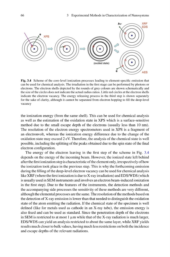
Fig. 3.4 Scheme of the core-level ionization processes leading to element-specific emission that
can be used for chemical analysis. The irradiation in the first stage can be performed by photons or
electrons. The electron shells depicted by the rounds of grey colours are shown schematically and
the size of the circles does not indicate the actual radius ratios. Little red circles at the electron shells
indicate the electron vacancy. The energy releasing process in the third step is shown separately
for the sake of clarity, although it cannot be separated from electron hopping to fill the deep-level
vacancy
the ionization energy (from the same shell). This can be used for chemical analysis
as well as the estimation of the oxidation state in XPS which is a surface-sensitive
method due to the small escape depth of the electrons (usually less than 10 nm).
The resolution of the electron energy spectrometers used in XPS is a fragment of
an electronvolt, whereas the ionization energy difference due to the change of the
oxidation state may exceed 2 eV. Therefore, the analysis of the chemical state is well
possible, including the splitting of the peaks obtained due to the spin state of the final
electron configuration.
The energy of the electron leaving in the first step of the scheme in Fig. 3.4
depends on the energy of the incoming beam. However, the ionized state left behind
after the first ionization step is characteristic of the element only, irrespectively of how
the ionization took place in the previous step. This is why the forthcoming emission
during the filling of the deep-level electron vacancy can be used for chemical analysis
like XRF (where the first ionization is due to X-ray irradiation) and EDS/WDS (which
is usually used in SEM instruments and involves an electron beam-induced ionization
in the first step). Due to the features of the instruments, the detection methods and
the accompanying side processes the sensitivity of these methods are very different,
although the elemental processes are the same. The resolution of the methods based on
the detection of X-ray emission is lower than that needed to distinguish the oxidation
state of the atom emitting the radiation. If the chemical state of the specimen is well
defined (like for metals used as cathode in an X-ray tube), the emission energy is
also fixed and can be used as standard. Since the penetration depth of the electrons
in SEM is restricted to at most 1 μm while that of the X-ray radiation is much larger,
EDS/WDS can yield an analysis restricted to about the same layer, while XRF yields
results much closer to bulk values, having much less restrictions on both the incidence
and escape depths of the relevant radiations.
3 Experimental Methods in Characterization of Nanosystems
Fig. 3.4 Scheme of the core-level ionization processes leading to element-specific emission that
can be used for chemical analysis. The irradiation in the first stage can be performed by photons or
electrons. The electron shells depicted by the rounds of grey colours are shown schematically and
the size of the circles does not indicate the actual radius ratios. Little red circles at the electron shells
indicate the electron vacancy. The energy releasing process in the third step is shown separately
for the sake of clarity, although it cannot be separated from electron hopping to fill the deep-level
vacancy
the ionization energy (from the same shell). This can be used for chemical analysis
as well as the estimation of the oxidation state in XPS which is a surface-sensitive
method due to the small escape depth of the electrons (usually less than 10 nm).
The resolution of the electron energy spectrometers used in XPS is a fragment of
an electronvolt, whereas the ionization energy difference due to the change of the
oxidation state may exceed 2 eV. Therefore, the analysis of the chemical state is well
possible, including the splitting of the peaks obtained due to the spin state of the final
electron configuration.
The energy of the electron leaving in the first step of the scheme in Fig. 3.4
depends on the energy of the incoming beam. However, the ionized state left behind
after the first ionization step is characteristic of the element only, irrespectively of how
the ionization took place in the previous step. This is why the forthcoming emission
during the filling of the deep-level electron vacancy can be used for chemical analysis
like XRF (where the first ionization is due to X-ray irradiation) and EDS/WDS (which
is usually used in SEM instruments and involves an electron beam-induced ionization
in the first step). Due to the features of the instruments, the detection methods and
the accompanying side processes the sensitivity of these methods are very different,
although the elemental processes are the same. The resolution of the methods based on
the detection of X-ray emission is lower than that needed to distinguish the oxidation
state of the atom emitting the radiation. If the chemical state of the specimen is well
defined (like for metals used as cathode in an X-ray tube), the emission energy is
also fixed and can be used as standard. Since the penetration depth of the electrons
in SEM is restricted to at most 1 μm while that of the X-ray radiation is much larger,
EDS/WDS can yield an analysis restricted to about the same layer, while XRF yields
results much closer to bulk values, having much less restrictions on both the incidence
and escape depths of the relevant radiations.