

58
3 Experimental Methods in Characterization of Nanosystems
Table 3.1
(continued)
Name of the method
(abbreviation, if any)
Range (wavelength or
photon energy)
Interaction of the beam with
the specimen
Signal detected (with some
details of the detection)
Information yield of the
method
X-ray photoelectron
spectroscopy (XPS)
X-ray
(1–2 keV)
Ionization of the atoms of a
solid material (in the near
surface zone) followed by
electron/X-ray emission
Flux of the electrons emitted
with various energies (out of
the incoming beam direction)
Surface composition,
oxidation state of the atoms
detected
X-ray fluorescence (XRF)
spectroscopy
X-ray (λ: 1.2–0.09 nm; up
to 60 keV)
Core-level ionization
followed by electron hopping
and emission of
characteristic X-ray
X-ray intensity as a function of
either energy or wavelength
(out of the incoming beam
direction)
Elemental composition
(insensitive to oxidation
state), for sodium and
elements of higher atomic
number
X-ray reflectometry
(small-angle X-ray scattering)
X-ray
Reflection at layer
boundaries
Radiation scattered elastically
in a small angle
Layer structure, interface
roughness
X-ray diffractometry (XRD)
X-ray (λ: 0.01–10 nm in
general; for metals:
0.07–0.18 nm)
Diffraction (interference of
beams elastically scattered
by atoms of identical crystal
planes)
X-ray (preferably of the same
wavelength, excluding
fluorescence)
Interplanar spacing of the
atoms in the direction of the
scattering vector, texture,
grain size (from peak
broadening)
X-ray absorption near-edge
structure (XANES)
X-ray (about 0.2–35 keV)
Absorption followed by
electron emission from the
core shells; interference of
the ejected and scattered
electron wave with the
incoming X-ray beam
Transmitted beam intensity as
a function of X-ray photon
energy near the absorption
edge (about 5 to 200 eV)
Oxidation state and
coordination environment of
the absorbing atom
(continued)
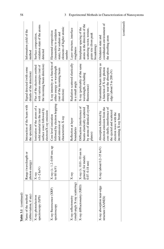
3 Experimental Methods in Characterization of Nanosystems
Table 3.1
(continued)
Name of the method
(abbreviation, if any)
Range (wavelength or
photon energy)
Interaction of the beam with
the specimen
Signal detected (with some
details of the detection)
Information yield of the
method
X-ray photoelectron
spectroscopy (XPS)
X-ray
(1–2 keV)
Ionization of the atoms of a
solid material (in the near
surface zone) followed by
electron/X-ray emission
Flux of the electrons emitted
with various energies (out of
the incoming beam direction)
Surface composition,
oxidation state of the atoms
detected
X-ray fluorescence (XRF)
spectroscopy
X-ray (λ: 1.2–0.09 nm; up
to 60 keV)
Core-level ionization
followed by electron hopping
and emission of
characteristic X-ray
X-ray intensity as a function of
either energy or wavelength
(out of the incoming beam
direction)
Elemental composition
(insensitive to oxidation
state), for sodium and
elements of higher atomic
number
X-ray reflectometry
(small-angle X-ray scattering)
X-ray
Reflection at layer
boundaries
Radiation scattered elastically
in a small angle
Layer structure, interface
roughness
X-ray diffractometry (XRD)
X-ray (λ: 0.01–10 nm in
general; for metals:
0.07–0.18 nm)
Diffraction (interference of
beams elastically scattered
by atoms of identical crystal
planes)
X-ray (preferably of the same
wavelength, excluding
fluorescence)
Interplanar spacing of the
atoms in the direction of the
scattering vector, texture,
grain size (from peak
broadening)
X-ray absorption near-edge
structure (XANES)
X-ray (about 0.2–35 keV)
Absorption followed by
electron emission from the
core shells; interference of
the ejected and scattered
electron wave with the
incoming X-ray beam
Transmitted beam intensity as
a function of X-ray photon
energy near the absorption
edge (about 5 to 200 eV)
Oxidation state and
coordination environment of
the absorbing atom
(continued)