

12.2 Two-Plate Thin-Layer Cells for Electron Microscopic …
435
possible, hence opening the way for the in situ identification of the phase and its
orientation.
A factor that is very unusual in macroscopic electrochemical experiments may
arise in all in situ TEM studies. Namely, the electron beam itself is capable of
generating or modifying chemical reactions. Here, we encounter something similar
to quantum mechanical observations where the probe method itself modifies the
system to be measured. Therefore, whenever it is possible, this has to be taken into
account and results separated from beam-induced effects should be given, unless the
chemical changes originating from the electron beam are exploited purposefully. The
latter method as a special field of electrochemistry will be dealt with in Sect. 12.2.4.
12.2.2 In Situ Electrochemical Nucleation Studies with TEM
The first fundamental study with in situ electrochemical nucleation used Cu deposition as a model system [31]. The solution concentration was 0.3 M, and the deposition
experiments were carried out with constant current density of either 5 or 50 mA cm
−2 .
The voltammogram in the thin-layer cell was equivalent to that obtained with a
conventional cell arrangement, the only difference being the impact of the uncompensated ohmic drop. It was carefully controlled that the surface inhomogeneity
of the polycrystalline Au substrate does not influence the nucleation in subsequent
experiments; i.e., nucleation sites in repetitive experiments with the same substrate
were uncorrelated. The result of the experiments was that the areal density of the Cu
grain grew steadily in the first period of about 2.5 s, but later practically no nucleation
event was observed. This is good agreement with what is generally thought about
nucleation. Namely, even if nucleation is progressive, the depletion of the media for
the reactant will increase the nucleation barrier so much that after a certain period
only grain growth can be observed.
After the proof-of-concept trial, the electrochemical manipulation was improved,
and in situ TEM observations were accompanied also by detailed electrochemical
data [32, 37, 38]. The quantitative analysis of the cluster radius r as a function of time
yielded the result that the nucleation and growth process can be divided into periods,
as indicated by the two subsequent linear sections in the log r vs log t function.
The exponents found experimentally for the start of the nucleus formation could be
correlated to the r ∝ t
1/2 law originating from the diffusion-limited growth of the
individual nuclei without the overlap of the hemispherical diffusion field around each
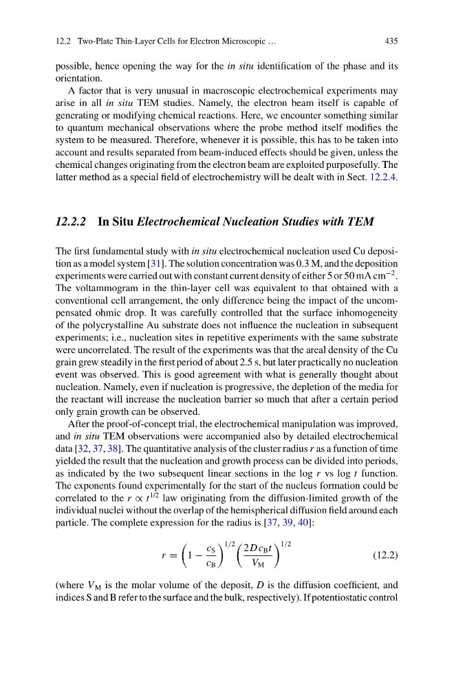
particle. The complete expression for the radius is [37, 39, 40]:
r =
1 −
c S
c B
1/2
2D c B t
V M
1/2
(12.2)
(where V M is the molar volume of the deposit, D is the diffusion coefficient, and
indices S and B refer to the surface and the bulk, respectively). If potentiostatic control
435
possible, hence opening the way for the in situ identification of the phase and its
orientation.
A factor that is very unusual in macroscopic electrochemical experiments may
arise in all in situ TEM studies. Namely, the electron beam itself is capable of
generating or modifying chemical reactions. Here, we encounter something similar
to quantum mechanical observations where the probe method itself modifies the
system to be measured. Therefore, whenever it is possible, this has to be taken into
account and results separated from beam-induced effects should be given, unless the
chemical changes originating from the electron beam are exploited purposefully. The
latter method as a special field of electrochemistry will be dealt with in Sect. 12.2.4.
12.2.2 In Situ Electrochemical Nucleation Studies with TEM
The first fundamental study with in situ electrochemical nucleation used Cu deposition as a model system [31]. The solution concentration was 0.3 M, and the deposition
experiments were carried out with constant current density of either 5 or 50 mA cm
−2 .
The voltammogram in the thin-layer cell was equivalent to that obtained with a
conventional cell arrangement, the only difference being the impact of the uncompensated ohmic drop. It was carefully controlled that the surface inhomogeneity
of the polycrystalline Au substrate does not influence the nucleation in subsequent
experiments; i.e., nucleation sites in repetitive experiments with the same substrate
were uncorrelated. The result of the experiments was that the areal density of the Cu
grain grew steadily in the first period of about 2.5 s, but later practically no nucleation
event was observed. This is good agreement with what is generally thought about
nucleation. Namely, even if nucleation is progressive, the depletion of the media for
the reactant will increase the nucleation barrier so much that after a certain period
only grain growth can be observed.
After the proof-of-concept trial, the electrochemical manipulation was improved,
and in situ TEM observations were accompanied also by detailed electrochemical
data [32, 37, 38]. The quantitative analysis of the cluster radius r as a function of time
yielded the result that the nucleation and growth process can be divided into periods,
as indicated by the two subsequent linear sections in the log r vs log t function.
The exponents found experimentally for the start of the nucleus formation could be
correlated to the r ∝ t
1/2 law originating from the diffusion-limited growth of the
individual nuclei without the overlap of the hemispherical diffusion field around each
particle. The complete expression for the radius is [37, 39, 40]:
r =
1 −
c S
c B
1/2
2D c B t
V M
1/2
(12.2)
(where V M is the molar volume of the deposit, D is the diffusion coefficient, and
indices S and B refer to the surface and the bulk, respectively). If potentiostatic control