

390
11 Templated Systems
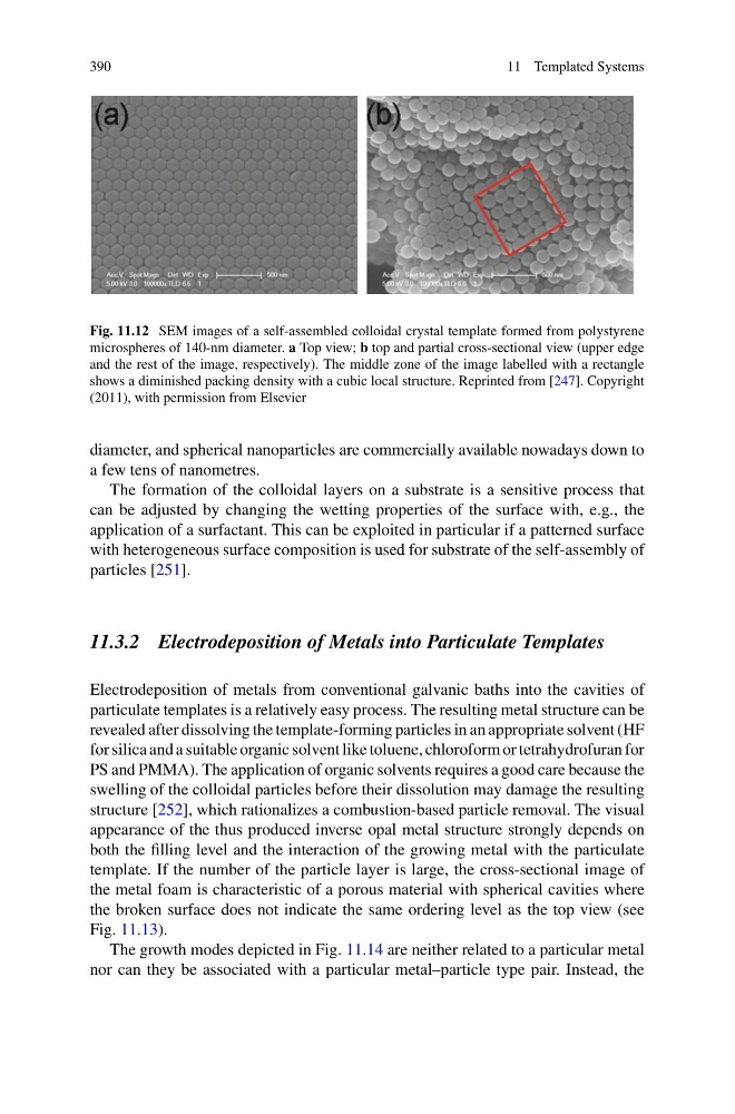
Fig. 11.12 SEM images of a self-assembled colloidal crystal template formed from polystyrene
microspheres of 140-nm diameter. a Top view; b top and partial cross-sectional view (upper edge
and the rest of the image, respectively). The middle zone of the image labelled with a rectangle
shows a diminished packing density with a cubic local structure. Reprinted from [247]. Copyright
(2011), with permission from Elsevier
diameter, and spherical nanoparticles are commercially available nowadays down to
a few tens of nanometres.
The formation of the colloidal layers on a substrate is a sensitive process that
can be adjusted by changing the wetting properties of the surface with, e.g., the
application of a surfactant. This can be exploited in particular if a patterned surface
with heterogeneous surface composition is used for substrate of the self-assembly of
particles [251].
11.3.2 Electrodeposition of Metals into Particulate Templates
Electrodeposition of metals from conventional galvanic baths into the cavities of
particulate templates is a relatively easy process. The resulting metal structure can be
revealed after dissolving the template-forming particles in an appropriate solvent (HF
for silica and a suitable organic solvent like toluene, chloroform or tetrahydrofuran for
PS and PMMA). The application of organic solvents requires a good care because the
swelling of the colloidal particles before their dissolution may damage the resulting
structure [252], which rationalizes a combustion-based particle removal. The visual
appearance of the thus produced inverse opal metal structure strongly depends on
both the filling level and the interaction of the growing metal with the particulate
template. If the number of the particle layer is large, the cross-sectional image of
the metal foam is characteristic of a porous material with spherical cavities where
the broken surface does not indicate the same ordering level as the top view (see
Fig. 11.13).
The growth modes depicted in Fig. 11.14 are neither related to a particular metal
nor can they be associated with a particular metal–particle type pair. Instead, the
11 Templated Systems
Fig. 11.12 SEM images of a self-assembled colloidal crystal template formed from polystyrene
microspheres of 140-nm diameter. a Top view; b top and partial cross-sectional view (upper edge
and the rest of the image, respectively). The middle zone of the image labelled with a rectangle
shows a diminished packing density with a cubic local structure. Reprinted from [247]. Copyright
(2011), with permission from Elsevier
diameter, and spherical nanoparticles are commercially available nowadays down to
a few tens of nanometres.
The formation of the colloidal layers on a substrate is a sensitive process that
can be adjusted by changing the wetting properties of the surface with, e.g., the
application of a surfactant. This can be exploited in particular if a patterned surface
with heterogeneous surface composition is used for substrate of the self-assembly of
particles [251].
11.3.2 Electrodeposition of Metals into Particulate Templates
Electrodeposition of metals from conventional galvanic baths into the cavities of
particulate templates is a relatively easy process. The resulting metal structure can be
revealed after dissolving the template-forming particles in an appropriate solvent (HF
for silica and a suitable organic solvent like toluene, chloroform or tetrahydrofuran for
PS and PMMA). The application of organic solvents requires a good care because the
swelling of the colloidal particles before their dissolution may damage the resulting
structure [252], which rationalizes a combustion-based particle removal. The visual
appearance of the thus produced inverse opal metal structure strongly depends on
both the filling level and the interaction of the growing metal with the particulate
template. If the number of the particle layer is large, the cross-sectional image of
the metal foam is characteristic of a porous material with spherical cavities where
the broken surface does not indicate the same ordering level as the top view (see
Fig. 11.13).
The growth modes depicted in Fig. 11.14 are neither related to a particular metal
nor can they be associated with a particular metal–particle type pair. Instead, the