

Biomechanical Evaluation of Medical Rehabilitation for a Patient …
261
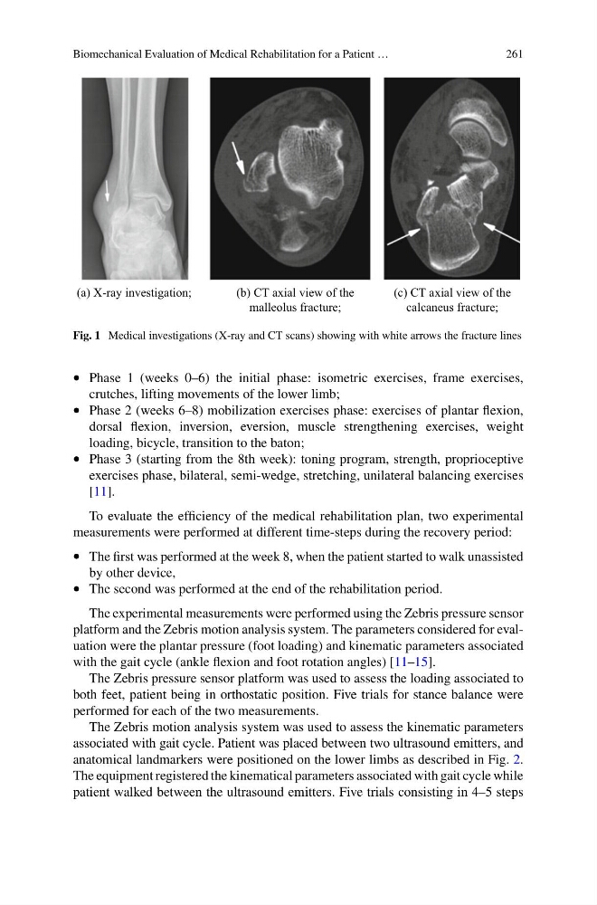
(a) X-ray investigation;
(b) CT axial view of the
malleolus fracture;
(c) CT axial view of the
calcaneus fracture;
Fig. 1 Medical investigations (X-ray and CT scans) showing with white arrows the fracture lines
• Phase 1 (weeks 0–6) the initial phase: isometric exercises, frame exercises,
crutches, lifting movements of the lower limb;
• Phase 2 (weeks 6–8) mobilization exercises phase: exercises of plantar flexion,
dorsal flexion, inversion, eversion, muscle strengthening exercises, weight
loading, bicycle, transition to the baton;
• Phase 3 (starting from the 8th week): toning program, strength, proprioceptive
exercises phase, bilateral, semi-wedge, stretching, unilateral balancing exercises
[11].
To evaluate the efficiency of the medical rehabilitation plan, two experimental
measurements were performed at different time-steps during the recovery period:
• The first was performed at the week 8, when the patient started to walk unassisted
by other device,
• The second was performed at the end of the rehabilitation period.
The experimental measurements were performed using the Zebris pressure sensor
platform and the Zebris motion analysis system. The parameters considered for evaluation were the plantar pressure (foot loading) and kinematic parameters associated
with the gait cycle (ankle flexion and foot rotation angles) [11–15].
The Zebris pressure sensor platform was used to assess the loading associated to
both feet, patient being in orthostatic position. Five trials for stance balance were
performed for each of the two measurements.
The Zebris motion analysis system was used to assess the kinematic parameters
associated with gait cycle. Patient was placed between two ultrasound emitters, and
anatomical landmarkers were positioned on the lower limbs as described in Fig. 2.
The equipment registered the kinematical parameters associated with gait cycle while
patient walked between the ultrasound emitters. Five trials consisting in 4–5 steps
261
(a) X-ray investigation;
(b) CT axial view of the
malleolus fracture;
(c) CT axial view of the
calcaneus fracture;
Fig. 1 Medical investigations (X-ray and CT scans) showing with white arrows the fracture lines
• Phase 1 (weeks 0–6) the initial phase: isometric exercises, frame exercises,
crutches, lifting movements of the lower limb;
• Phase 2 (weeks 6–8) mobilization exercises phase: exercises of plantar flexion,
dorsal flexion, inversion, eversion, muscle strengthening exercises, weight
loading, bicycle, transition to the baton;
• Phase 3 (starting from the 8th week): toning program, strength, proprioceptive
exercises phase, bilateral, semi-wedge, stretching, unilateral balancing exercises
[11].
To evaluate the efficiency of the medical rehabilitation plan, two experimental
measurements were performed at different time-steps during the recovery period:
• The first was performed at the week 8, when the patient started to walk unassisted
by other device,
• The second was performed at the end of the rehabilitation period.
The experimental measurements were performed using the Zebris pressure sensor
platform and the Zebris motion analysis system. The parameters considered for evaluation were the plantar pressure (foot loading) and kinematic parameters associated
with the gait cycle (ankle flexion and foot rotation angles) [11–15].
The Zebris pressure sensor platform was used to assess the loading associated to
both feet, patient being in orthostatic position. Five trials for stance balance were
performed for each of the two measurements.
The Zebris motion analysis system was used to assess the kinematic parameters
associated with gait cycle. Patient was placed between two ultrasound emitters, and
anatomical landmarkers were positioned on the lower limbs as described in Fig. 2.
The equipment registered the kinematical parameters associated with gait cycle while
patient walked between the ultrasound emitters. Five trials consisting in 4–5 steps