

32
crow flies,” and calculate length using ~2.5 Å per amino acid. For
POIs in which the line-of-sight between termini is blocked by
structure, we use a figure of <2.0 Å per amino acid to account for
the arc that the linker must take over the curved surface of the
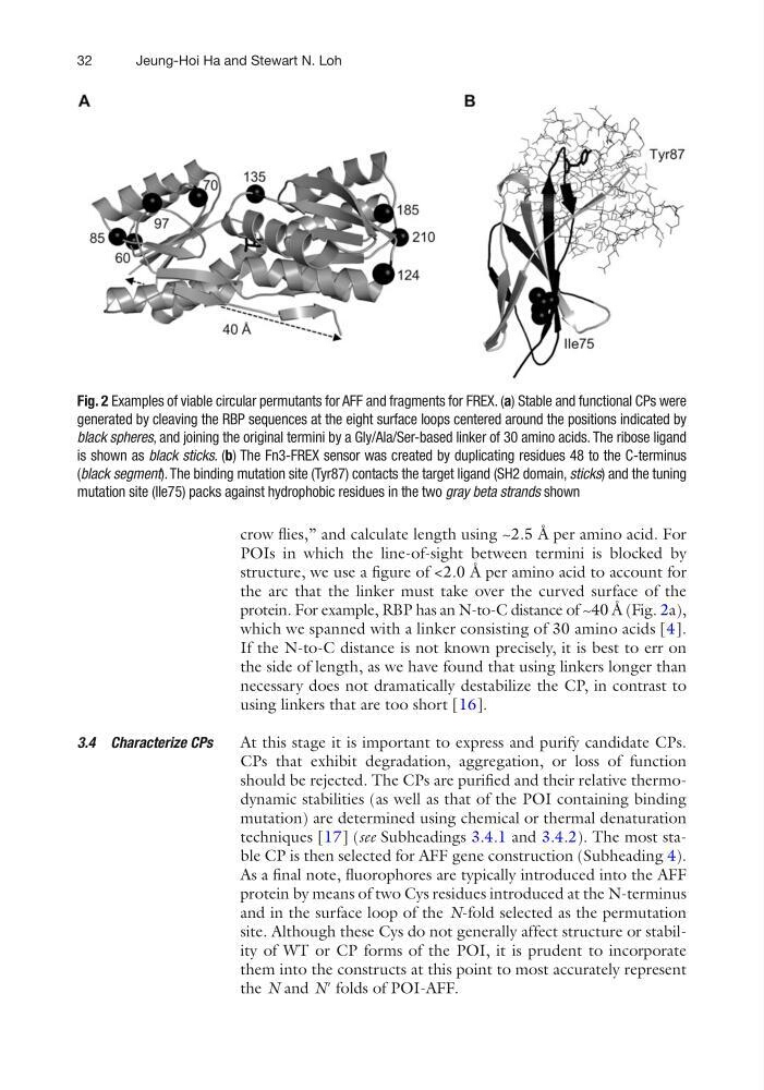
protein. For example, RBP has an N-to-C distance of ~40 Å (Fig. 2a),
which we spanned with a linker consisting of 30 amino acids [4].
If the N-to-C distance is not known precisely, it is best to err on
the side of length, as we have found that using linkers longer than
necessary does not dramatically destabilize the CP, in contrast to
using linkers that are too short [16].
At this stage it is important to express and purify candidate CPs.
CPs that exhibit degradation, aggregation, or loss of function
should be rejected. The CPs are purified and their relative thermodynamic stabilities (as well as that of the POI containing binding
mutation) are determined using chemical or thermal denaturation
techniques [17] (see Subheadings 3.4.1 and 3.4.2). The most stable CP is then selected for AFF gene construction (Subheading 4).
As a final note, fluorophores are typically introduced into the AFF
protein by means of two Cys residues introduced at the N-terminus
and in the surface loop of the N-fold selected as the permutation
site. Although these Cys do not generally affect structure or stability of WT or CP forms of the POI, it is prudent to incorporate
them into the constructs at this point to most accurately represent
the N and N′ folds of POI-AFF.
3.4 Characterize CPs
Fig. 2 Examples of viable circular permutants for AFF and fragments for FREX. (a) Stable and functional CPs were
generated by cleaving the RBP sequences at the eight surface loops centered around the positions indicated by
black spheres, and joining the original termini by a Gly/Ala/Ser-based linker of 30 amino acids. The ribose ligand
is shown as black sticks. (b) The Fn3-FREX sensor was created by duplicating residues 48 to the C-terminus
(black segment). The binding mutation site (Tyr87) contacts the target ligand (SH2 domain, sticks) and the tuning
mutation site (Ile75) packs against hydrophobic residues in the two gray beta strands shown
Jeung-Hoi Ha and Stewart N. Loh
crow flies,” and calculate length using ~2.5 Å per amino acid. For
POIs in which the line-of-sight between termini is blocked by
structure, we use a figure of <2.0 Å per amino acid to account for
the arc that the linker must take over the curved surface of the
protein. For example, RBP has an N-to-C distance of ~40 Å (Fig. 2a),
which we spanned with a linker consisting of 30 amino acids [4].
If the N-to-C distance is not known precisely, it is best to err on
the side of length, as we have found that using linkers longer than
necessary does not dramatically destabilize the CP, in contrast to
using linkers that are too short [16].
At this stage it is important to express and purify candidate CPs.
CPs that exhibit degradation, aggregation, or loss of function
should be rejected. The CPs are purified and their relative thermodynamic stabilities (as well as that of the POI containing binding
mutation) are determined using chemical or thermal denaturation
techniques [17] (see Subheadings 3.4.1 and 3.4.2). The most stable CP is then selected for AFF gene construction (Subheading 4).
As a final note, fluorophores are typically introduced into the AFF
protein by means of two Cys residues introduced at the N-terminus
and in the surface loop of the N-fold selected as the permutation
site. Although these Cys do not generally affect structure or stability of WT or CP forms of the POI, it is prudent to incorporate
them into the constructs at this point to most accurately represent
the N and N′ folds of POI-AFF.
3.4 Characterize CPs
Fig. 2 Examples of viable circular permutants for AFF and fragments for FREX. (a) Stable and functional CPs were
generated by cleaving the RBP sequences at the eight surface loops centered around the positions indicated by
black spheres, and joining the original termini by a Gly/Ala/Ser-based linker of 30 amino acids. The ribose ligand
is shown as black sticks. (b) The Fn3-FREX sensor was created by duplicating residues 48 to the C-terminus
(black segment). The binding mutation site (Tyr87) contacts the target ligand (SH2 domain, sticks) and the tuning
mutation site (Ile75) packs against hydrophobic residues in the two gray beta strands shown
Jeung-Hoi Ha and Stewart N. Loh