

310
radioactivity-based assay, which remains a gold standard for activity
measurements. The in vitro procedure can be completed in 9–10 h,
which allows for rapid and practical iterative optimization of a particular split kinase design. We have successfully used this approach
to construct split kinases for the catalytic domains of tyrosine
kinases Src, Lyn, Fyn, Abl, Fak, Hck, the catalytic domain of serine/threonine kinase PKA, and full-length tyrosine kinases Src,
Lyn, and Abl. We envision that other desired split enzymes can be
similarly designed, interrogated, and optimized.
The following protocol describes strategies used to construct a
split protein sensor for specific kinases, and to test the design by
expressing the fragments either using in vitro or in cellulo
approaches, and subsequently measuring kinase activity in its on
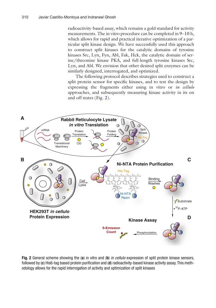
and off states (Fig. 2).
Ni-NTA Protein Purification
P-ATP
32
Substrate
HO
Phosphorylation
Ni
NTA
HO
Ni
NTA
His-Tag
Ni
NTA
Ni
NTA
Ni-NTA
Resin
ß-Emission
Count
Binding
Washes
Kinase Assay
Translational
Machinery
Protein
Translation
mRNA
Protein
Folding
Rabbit Reticulocyte Lysate
in vitro Translation
Split
Kinase
His-Tag
CID
HEK293T in cellulo
Protein Expression
s
Substrat Subs
HO O
Ni
NTA
HO HO O O
Ni
NTA
Ni
NTA
Ni-NT
Resi
Ni
NTA
TA
n
e
g
Ni NTA Pro
Split
nase
g
n
-Tag
s
S
Ki
His
in
ng
cellulo
K293T in c
in c
A
B
C
D
Cell
Lysis
Fig. 2 General scheme showing the (a) in vitro and (b) in cellulo expression of split protein kinase sensors,
followed by (c) His6-tag based protein purification and (d) radioactivity-based kinase activity assay. This methodology allows for the rapid interrogation of activity and optimization of split kinases
Javier Castillo-Montoya and Indraneel Ghosh
radioactivity-based assay, which remains a gold standard for activity
measurements. The in vitro procedure can be completed in 9–10 h,
which allows for rapid and practical iterative optimization of a particular split kinase design. We have successfully used this approach
to construct split kinases for the catalytic domains of tyrosine
kinases Src, Lyn, Fyn, Abl, Fak, Hck, the catalytic domain of serine/threonine kinase PKA, and full-length tyrosine kinases Src,
Lyn, and Abl. We envision that other desired split enzymes can be
similarly designed, interrogated, and optimized.
The following protocol describes strategies used to construct a
split protein sensor for specific kinases, and to test the design by
expressing the fragments either using in vitro or in cellulo
approaches, and subsequently measuring kinase activity in its on
and off states (Fig. 2).
Ni-NTA Protein Purification
P-ATP
32
Substrate
HO
Phosphorylation
Ni
NTA
HO
Ni
NTA
His-Tag
Ni
NTA
Ni
NTA
Ni-NTA
Resin
ß-Emission
Count
Binding
Washes
Kinase Assay
Translational
Machinery
Protein
Translation
mRNA
Protein
Folding
Rabbit Reticulocyte Lysate
in vitro Translation
Split
Kinase
His-Tag
CID
HEK293T in cellulo
Protein Expression
s
Substrat Subs
HO O
Ni
NTA
HO HO O O
Ni
NTA
Ni
NTA
Ni-NT
Resi
Ni
NTA
TA
n
e
g
Ni NTA Pro
Split
nase
g
n
-Tag
s
S
Ki
His
in
ng
cellulo
K293T in c
in c
A
B
C
D
Cell
Lysis
Fig. 2 General scheme showing the (a) in vitro and (b) in cellulo expression of split protein kinase sensors,
followed by (c) His6-tag based protein purification and (d) radioactivity-based kinase activity assay. This methodology allows for the rapid interrogation of activity and optimization of split kinases
Javier Castillo-Montoya and Indraneel Ghosh