

243
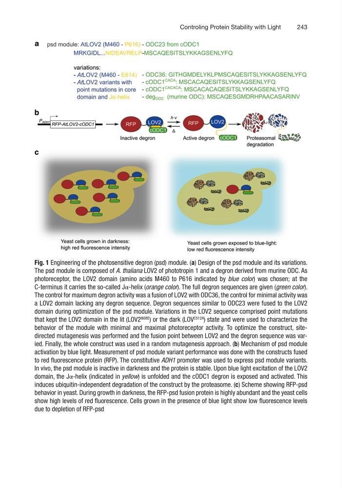
Fig. 1 Engineering of the photosensitive degron (psd) module. (a) Design of the psd module and its variations.
The psd module is composed of A. thaliana LOV2 of phototropin 1 and a degron derived from murine ODC. As
photoreceptor, the LOV2 domain (amino acids M460 to P616 indicated by blue color) was chosen; at the
C-terminus it carries the so-called Jα-helix (orange color). The full degron sequences are given (green color).
The control for maximum degron activity was a fusion of LOV2 with ODC36, the control for minimal activity was
a LOV2 domain lacking any degron sequence. Degron sequences similar to ODC23 were fused to the LOV2
domain during optimization of the psd module. Variations in the LOV2 sequence comprised point mutations
that kept the LOV2 domain in the lit (LOV2
I608E
) or the dark (LOV
C512A
) state and were used to characterize the
behavior of the module with minimal and maximal photoreceptor activity. To optimize the construct, sitedirected mutagenesis was performed and the fusion point between LOV2 and the degron sequence was varied. Finally, the whole construct was used in a random mutagenesis approach. (b) Mechanism of psd module
activation by blue light. Measurement of psd module variant performance was done with the constructs fused
to red fluorescence protein (RFP). The constitutive ADH1 promoter was used to express psd module variants.
In vivo, the psd module is inactive in darkness and the protein is stable. Upon blue light excitation of the LOV2
domain, the Jα-helix (indicated in yellow) is unfolded and the cODC1 degron is exposed and activated. This
induces ubiquitin-independent degradation of the construct by the proteasome. (c) Scheme showing RFP-psd
behavior in yeast. During growth in darkness, the RFP-psd fusion protein is highly abundant and the yeast cells
show high levels of red fluorescence. Cells grown in the presence of blue light show low fluorescence levels
due to depletion of RFP-psd
Controling Protein Stability with Light
Fig. 1 Engineering of the photosensitive degron (psd) module. (a) Design of the psd module and its variations.
The psd module is composed of A. thaliana LOV2 of phototropin 1 and a degron derived from murine ODC. As
photoreceptor, the LOV2 domain (amino acids M460 to P616 indicated by blue color) was chosen; at the
C-terminus it carries the so-called Jα-helix (orange color). The full degron sequences are given (green color).
The control for maximum degron activity was a fusion of LOV2 with ODC36, the control for minimal activity was
a LOV2 domain lacking any degron sequence. Degron sequences similar to ODC23 were fused to the LOV2
domain during optimization of the psd module. Variations in the LOV2 sequence comprised point mutations
that kept the LOV2 domain in the lit (LOV2
I608E
) or the dark (LOV
C512A
) state and were used to characterize the
behavior of the module with minimal and maximal photoreceptor activity. To optimize the construct, sitedirected mutagenesis was performed and the fusion point between LOV2 and the degron sequence was varied. Finally, the whole construct was used in a random mutagenesis approach. (b) Mechanism of psd module
activation by blue light. Measurement of psd module variant performance was done with the constructs fused
to red fluorescence protein (RFP). The constitutive ADH1 promoter was used to express psd module variants.
In vivo, the psd module is inactive in darkness and the protein is stable. Upon blue light excitation of the LOV2
domain, the Jα-helix (indicated in yellow) is unfolded and the cODC1 degron is exposed and activated. This
induces ubiquitin-independent degradation of the construct by the proteasome. (c) Scheme showing RFP-psd
behavior in yeast. During growth in darkness, the RFP-psd fusion protein is highly abundant and the yeast cells
show high levels of red fluorescence. Cells grown in the presence of blue light show low fluorescence levels
due to depletion of RFP-psd
Controling Protein Stability with Light