

225
interaction controls. Therefore, it is strongly advisable to include
positive controls (i.e., proteins that do interact with the candidate
of interest) and negative controls (i.e., proteins that do not bind
to the candidate of interest) for each assay designed.
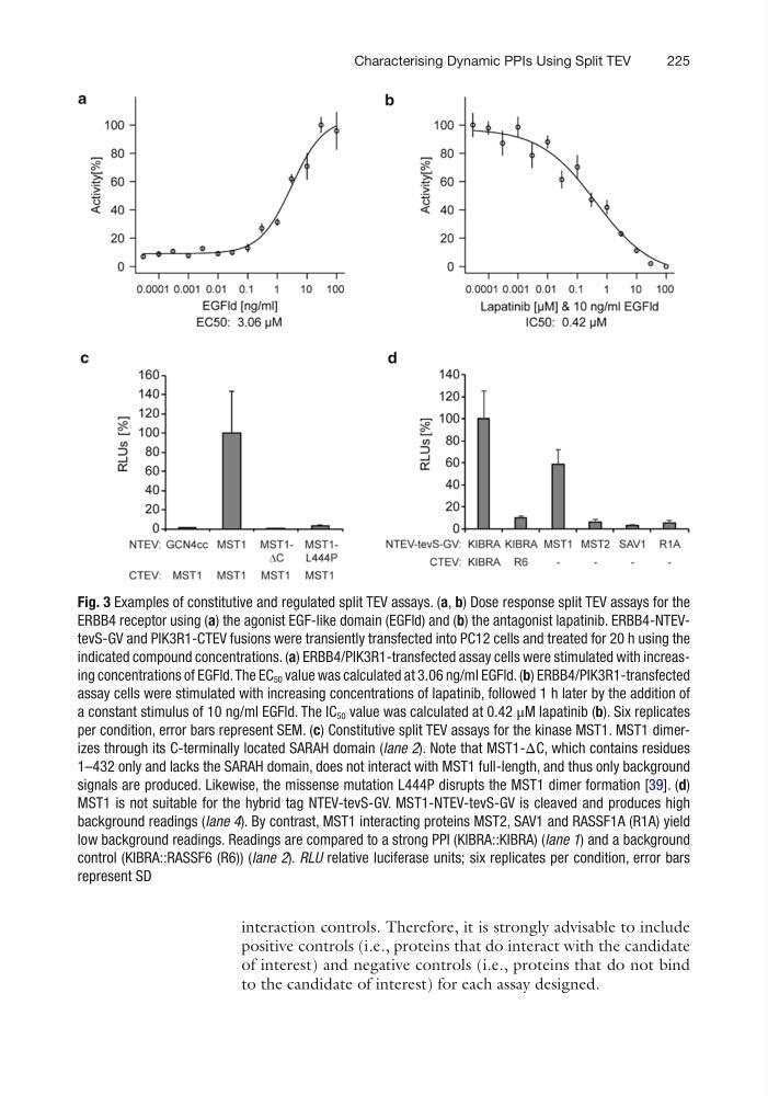
Fig. 3 Examples of constitutive and regulated split TEV assays. (a, b) Dose response split TEV assays for the
ERBB4 receptor using (a) the agonist EGF-like domain (EGFld) and (b) the antagonist lapatinib. ERBB4-NTEVtevS-GV and PIK3R1-CTEV fusions were transiently transfected into PC12 cells and treated for 20 h using the
indicated compound concentrations. (a) ERBB4/PIK3R1-transfected assay cells were stimulated with increasing concentrations of EGFld. The EC 50 value was calculated at 3.06 ng/ml EGFld. (b) ERBB4/PIK3R1-transfected
assay cells were stimulated with increasing concentrations of lapatinib, followed 1 h later by the addition of
a constant stimulus of 10 ng/ml EGFld. The IC 50 value was calculated at 0.42 μM lapatinib (b). Six replicates
per condition, error bars represent SEM. (c) Constitutive split TEV assays for the kinase MST1. MST1 dimerizes through its C-terminally located SARAH domain (lane 2). Note that MST1-ΔC, which contains residues
1–432 only and lacks the SARAH domain, does not interact with MST1 full-length, and thus only background
signals are produced. Likewise, the missense mutation L444P disrupts the MST1 dimer formation [39]. (d)
MST1 is not suitable for the hybrid tag NTEV-tevS-GV. MST1-NTEV-tevS-GV is cleaved and produces high
background readings (lane 4). By contrast, MST1 interacting proteins MST2, SAV1 and RASSF1A (R1A) yield
low background readings. Readings are compared to a strong PPI (KIBRA::KIBRA) (lane 1) and a background
control (KIBRA::RASSF6 (R6)) (lane 2). RLU relative luciferase units; six replicates per condition, error bars
represent SD
Characterising Dynamic PPIs Using Split TEV
interaction controls. Therefore, it is strongly advisable to include
positive controls (i.e., proteins that do interact with the candidate
of interest) and negative controls (i.e., proteins that do not bind
to the candidate of interest) for each assay designed.
Fig. 3 Examples of constitutive and regulated split TEV assays. (a, b) Dose response split TEV assays for the
ERBB4 receptor using (a) the agonist EGF-like domain (EGFld) and (b) the antagonist lapatinib. ERBB4-NTEVtevS-GV and PIK3R1-CTEV fusions were transiently transfected into PC12 cells and treated for 20 h using the
indicated compound concentrations. (a) ERBB4/PIK3R1-transfected assay cells were stimulated with increasing concentrations of EGFld. The EC 50 value was calculated at 3.06 ng/ml EGFld. (b) ERBB4/PIK3R1-transfected
assay cells were stimulated with increasing concentrations of lapatinib, followed 1 h later by the addition of
a constant stimulus of 10 ng/ml EGFld. The IC 50 value was calculated at 0.42 μM lapatinib (b). Six replicates
per condition, error bars represent SEM. (c) Constitutive split TEV assays for the kinase MST1. MST1 dimerizes through its C-terminally located SARAH domain (lane 2). Note that MST1-ΔC, which contains residues
1–432 only and lacks the SARAH domain, does not interact with MST1 full-length, and thus only background
signals are produced. Likewise, the missense mutation L444P disrupts the MST1 dimer formation [39]. (d)
MST1 is not suitable for the hybrid tag NTEV-tevS-GV. MST1-NTEV-tevS-GV is cleaved and produces high
background readings (lane 4). By contrast, MST1 interacting proteins MST2, SAV1 and RASSF1A (R1A) yield
low background readings. Readings are compared to a strong PPI (KIBRA::KIBRA) (lane 1) and a background
control (KIBRA::RASSF6 (R6)) (lane 2). RLU relative luciferase units; six replicates per condition, error bars
represent SD
Characterising Dynamic PPIs Using Split TEV