

223
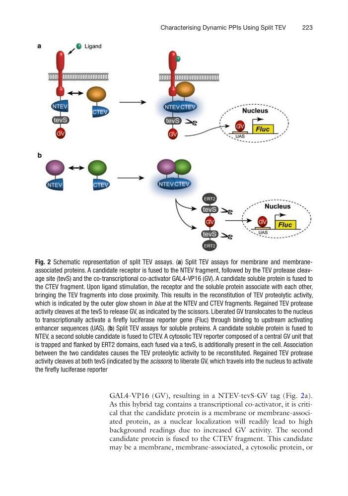
GAL4-VP16 (GV), resulting in a NTEV-tevS-GV tag (Fig. 2a).
As this hybrid tag contains a transcriptional co-activator, it is critical that the candidate protein is a membrane or membrane-associated protein, as a nuclear localization will readily lead to high
background readings due to increased GV activity. The second
candidate protein is fused to the CTEV fragment. This candidate
may be a membrane, membrane-associated, a cytosolic protein, or
Fig. 2 Schematic representation of split TEV assays. (a) Split TEV assays for membrane and membraneassociated proteins. A candidate receptor is fused to the NTEV fragment, followed by the TEV protease cleavage site (tevS) and the co-transcriptional co-activator GAL4-VP16 (GV). A candidate soluble protein is fused to
the CTEV fragment. Upon ligand stimulation, the receptor and the soluble protein associate with each other,
bringing the TEV fragments into close proximity. This results in the reconstitution of TEV proteolytic activity,
which is indicated by the outer glow shown in blue at the NTEV and CTEV fragments. Regained TEV protease
activity cleaves at the tevS to release GV, as indicated by the scissors. Liberated GV translocates to the nucleus
to transcriptionally activate a firefly luciferase reporter gene (Fluc) through binding to upstream activating
enhancer sequences (UAS). (b) Split TEV assays for soluble proteins. A candidate soluble protein is fused to
NTEV, a second soluble candidate is fused to CTEV. A cytosolic TEV reporter composed of a central GV unit that
is trapped and flanked by ERT2 domains, each fused via a tevS, is additionally present in the cell. Association
between the two candidates causes the TEV proteolytic activity to be reconstituted. Regained TEV protease
activity cleaves at both tevS (indicated by the scissors) to liberate GV, which travels into the nucleus to activate
the firefly luciferase reporter
Characterising Dynamic PPIs Using Split TEV
GAL4-VP16 (GV), resulting in a NTEV-tevS-GV tag (Fig. 2a).
As this hybrid tag contains a transcriptional co-activator, it is critical that the candidate protein is a membrane or membrane-associated protein, as a nuclear localization will readily lead to high
background readings due to increased GV activity. The second
candidate protein is fused to the CTEV fragment. This candidate
may be a membrane, membrane-associated, a cytosolic protein, or
Fig. 2 Schematic representation of split TEV assays. (a) Split TEV assays for membrane and membraneassociated proteins. A candidate receptor is fused to the NTEV fragment, followed by the TEV protease cleavage site (tevS) and the co-transcriptional co-activator GAL4-VP16 (GV). A candidate soluble protein is fused to
the CTEV fragment. Upon ligand stimulation, the receptor and the soluble protein associate with each other,
bringing the TEV fragments into close proximity. This results in the reconstitution of TEV proteolytic activity,
which is indicated by the outer glow shown in blue at the NTEV and CTEV fragments. Regained TEV protease
activity cleaves at the tevS to release GV, as indicated by the scissors. Liberated GV translocates to the nucleus
to transcriptionally activate a firefly luciferase reporter gene (Fluc) through binding to upstream activating
enhancer sequences (UAS). (b) Split TEV assays for soluble proteins. A candidate soluble protein is fused to
NTEV, a second soluble candidate is fused to CTEV. A cytosolic TEV reporter composed of a central GV unit that
is trapped and flanked by ERT2 domains, each fused via a tevS, is additionally present in the cell. Association
between the two candidates causes the TEV proteolytic activity to be reconstituted. Regained TEV protease
activity cleaves at both tevS (indicated by the scissors) to liberate GV, which travels into the nucleus to activate
the firefly luciferase reporter
Characterising Dynamic PPIs Using Split TEV