

222
blocks the active site of the TEV protease [37]. NTEV and CTEV
fragments are fused to candidate proteins, preferably to the
C-terminal end of the protein candidates. For NTEV, N-terminal
fusions are also functional. Protein Interaction-induced reassembly
of the NTEV and CTEV moieties leads to the reconstitution of
TEV proteolytic activity, which activates TEV-specific reporters.
These can be either of proteolytic or of transcriptional type, and
they maybe fluorescent or luminescent-based. Notably, the
luciferase- based transcriptional reporters proved to be most sensitive and robust as the readout is functionally uncoupled from the
interaction event itself and background readings were commonly
lower compared to other options. Transcriptional reporters based
on luciferase exhibit another feature as they comprise three levels
of signal amplification: In addition to the proteolytic cleavage and
a transcriptional amplification step, enzyme-based (i.e., luciferase)
reporters allow a third step of amplifying the initial signal resulting
in a robust and sensitive readout.
For monitoring dynamic PPIs at the membrane, a membrane protein or membrane-associated protein candidate is fused to the
NTEV fragment along with a TEV protease cleavage site (tevS,
encoded by the amino acids ENLYFQ’G; the TEV protease cleaves
between Q and G) and the artificial transcriptional co-activator
1.2 Split TEV Assays
for Membrane
and MembraneAssociated Proteins
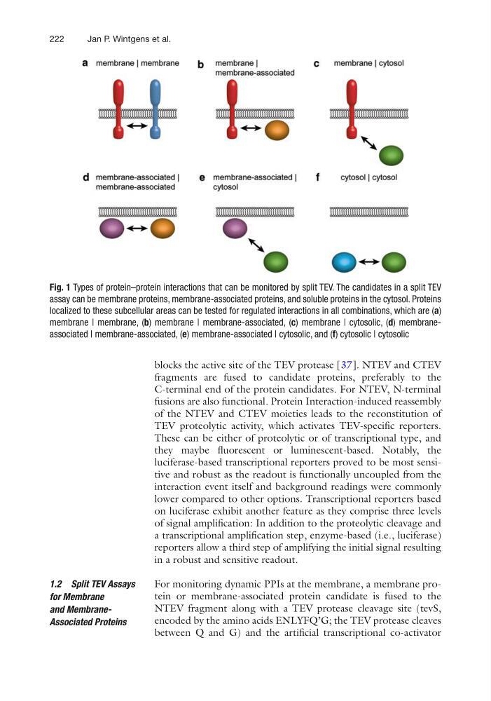
Fig. 1 Types of protein–protein interactions that can be monitored by split TEV. The candidates in a split TEV
assay can be membrane proteins, membrane-associated proteins, and soluble proteins in the cytosol. Proteins
localized to these subcellular areas can be tested for regulated interactions in all combinations, which are (a)
membrane | membrane, (b) membrane | membrane-associated, (c) membrane | cytosolic, (d) membraneassociated | membrane-associated, (e) membrane-associated | cytosolic, and (f) cytosolic | cytosolic
Jan P. Wintgens et al.
blocks the active site of the TEV protease [37]. NTEV and CTEV
fragments are fused to candidate proteins, preferably to the
C-terminal end of the protein candidates. For NTEV, N-terminal
fusions are also functional. Protein Interaction-induced reassembly
of the NTEV and CTEV moieties leads to the reconstitution of
TEV proteolytic activity, which activates TEV-specific reporters.
These can be either of proteolytic or of transcriptional type, and
they maybe fluorescent or luminescent-based. Notably, the
luciferase- based transcriptional reporters proved to be most sensitive and robust as the readout is functionally uncoupled from the
interaction event itself and background readings were commonly
lower compared to other options. Transcriptional reporters based
on luciferase exhibit another feature as they comprise three levels
of signal amplification: In addition to the proteolytic cleavage and
a transcriptional amplification step, enzyme-based (i.e., luciferase)
reporters allow a third step of amplifying the initial signal resulting
in a robust and sensitive readout.
For monitoring dynamic PPIs at the membrane, a membrane protein or membrane-associated protein candidate is fused to the
NTEV fragment along with a TEV protease cleavage site (tevS,
encoded by the amino acids ENLYFQ’G; the TEV protease cleaves
between Q and G) and the artificial transcriptional co-activator
1.2 Split TEV Assays
for Membrane
and MembraneAssociated Proteins
Fig. 1 Types of protein–protein interactions that can be monitored by split TEV. The candidates in a split TEV
assay can be membrane proteins, membrane-associated proteins, and soluble proteins in the cytosol. Proteins
localized to these subcellular areas can be tested for regulated interactions in all combinations, which are (a)
membrane | membrane, (b) membrane | membrane-associated, (c) membrane | cytosolic, (d) membraneassociated | membrane-associated, (e) membrane-associated | cytosolic, and (f) cytosolic | cytosolic
Jan P. Wintgens et al.