

191
6. Derive the hydrolysis rate by calculating the slope of the absorbance in time of the initial, linear regime. Correct for background
hydrolysis of nitrocefin in the absence of enzyme (Fig. 5).
1. Mix ODN1-TEM1-β-lactamase, ODN2-BLIP, and ssDNA
template (with a target recognition sequence length of 40 nucleotides) in PBS
+
at low micromolar concentrations in a ratio of
1:2:1.2 and incubate for 30 min at room temperature.
2. Dilute the mixture in PBS
+
to a final concentration of 5 nM
ODN1-TEM1-β-lactamase.
3. In a 96-well plate, mix 4 μL of the diluted complex with
152 μL PBS
+
and 24 μL target oligonucleotide (concentrations
ranging from 100 pM to 20 nM) and incubate for 1 h at room
temperature.
4. Add 20 μL CCF2-FA (20 μM in PBS), yielding a final concentration of 100 pM DNA-directed enzyme-inhibitor complex,
10 pM to 2 nM target oligonucleotide, and 2 μM CCF2- FA.
Incubate for 45 min at room temperature.
3.6 Enzyme Activity
Assay at Subnanomolar Input
Oligonucleotide
Concentrations
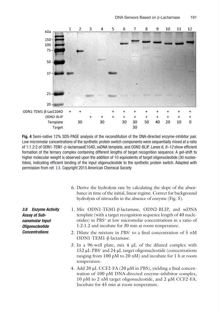
Fig. 4 Semi-native 12% SDS-PAGE analysis of the reconstitution of the DNA-directed enzyme-inhibitor pair.
Low micromolar concentrations of the synthetic protein switch components were sequentially mixed at a ratio
of 1:1.2:2 of ODN1-TEM1-β-lactamaseE104D, ssDNA template, and ODN2-BLIP. Lanes 6, 8–12 show efficient
formation of the ternary complex containing different lengths of target recognition sequence. A gel-shift to
higher molecular weight is observed upon the addition of 10 equivalents of target oligonucleotide (30 nucleotides), indicating efficient binding of the input oligonucleotide to the synthetic protein switch. Adapted with
permission from ref. 13. Copyright 2015 American Chemical Society
DNA Sensors Based on β-Lactamase
6. Derive the hydrolysis rate by calculating the slope of the absorbance in time of the initial, linear regime. Correct for background
hydrolysis of nitrocefin in the absence of enzyme (Fig. 5).
1. Mix ODN1-TEM1-β-lactamase, ODN2-BLIP, and ssDNA
template (with a target recognition sequence length of 40 nucleotides) in PBS
+
at low micromolar concentrations in a ratio of
1:2:1.2 and incubate for 30 min at room temperature.
2. Dilute the mixture in PBS
+
to a final concentration of 5 nM
ODN1-TEM1-β-lactamase.
3. In a 96-well plate, mix 4 μL of the diluted complex with
152 μL PBS
+
and 24 μL target oligonucleotide (concentrations
ranging from 100 pM to 20 nM) and incubate for 1 h at room
temperature.
4. Add 20 μL CCF2-FA (20 μM in PBS), yielding a final concentration of 100 pM DNA-directed enzyme-inhibitor complex,
10 pM to 2 nM target oligonucleotide, and 2 μM CCF2- FA.
Incubate for 45 min at room temperature.
3.6 Enzyme Activity
Assay at Subnanomolar Input
Oligonucleotide
Concentrations
Fig. 4 Semi-native 12% SDS-PAGE analysis of the reconstitution of the DNA-directed enzyme-inhibitor pair.
Low micromolar concentrations of the synthetic protein switch components were sequentially mixed at a ratio
of 1:1.2:2 of ODN1-TEM1-β-lactamaseE104D, ssDNA template, and ODN2-BLIP. Lanes 6, 8–12 show efficient
formation of the ternary complex containing different lengths of target recognition sequence. A gel-shift to
higher molecular weight is observed upon the addition of 10 equivalents of target oligonucleotide (30 nucleotides), indicating efficient binding of the input oligonucleotide to the synthetic protein switch. Adapted with
permission from ref. 13. Copyright 2015 American Chemical Society
DNA Sensors Based on β-Lactamase