

102
can be used to give a quantitative output. However, many binding
proteins do not undergo a sufficiently large conformational change
when they bind to the analyte of interest. For these binding proteins, modular biosensors can be used to give larger artificial conformational changes (see [8], and references therein). Our
laboratory has introduced two such examples: SNAP-tag-based
indicators with a Fluorescent Intramolecular Tether (SNIFITs)
and LUCiferase-based Indicators of Drugs (LUCIDs).
SNIFITs and LUCIDs are semisynthetic sensors composed of
(1) a recombinant fusion protein and (2) a synthetic intramolecular
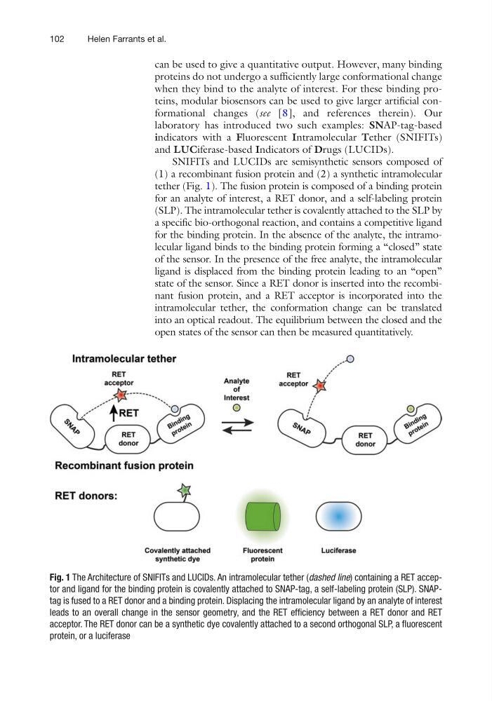
tether (Fig. 1). The fusion protein is composed of a binding protein
for an analyte of interest, a RET donor, and a self-labeling protein
(SLP). The intramolecular tether is covalently attached to the SLP by
a specific bio-orthogonal reaction, and contains a competitive ligand
for the binding protein. In the absence of the analyte, the intramolecular ligand binds to the binding protein forming a “closed” state
of the sensor. In the presence of the free analyte, the intramolecular
ligand is displaced from the binding protein leading to an “open”
state of the sensor. Since a RET donor is inserted into the recombinant fusion protein, and a RET acceptor is incorporated into the
intramolecular tether, the conformation change can be translated
into an optical readout. The equilibrium between the closed and the
open states of the sensor can then be measured quantitatively.
Fig. 1 The Architecture of SNIFITs and LUCIDs. An intramolecular tether (dashed line) containing a RET acceptor and ligand for the binding protein is covalently attached to SNAP-tag, a self-labeling protein (SLP). SNAPtag is fused to a RET donor and a binding protein. Displacing the intramolecular ligand by an analyte of interest
leads to an overall change in the sensor geometry, and the RET efficiency between a RET donor and RET
acceptor. The RET donor can be a synthetic dye covalently attached to a second orthogonal SLP, a fluorescent
protein, or a luciferase
Helen Farrants et al.
can be used to give a quantitative output. However, many binding
proteins do not undergo a sufficiently large conformational change
when they bind to the analyte of interest. For these binding proteins, modular biosensors can be used to give larger artificial conformational changes (see [8], and references therein). Our
laboratory has introduced two such examples: SNAP-tag-based
indicators with a Fluorescent Intramolecular Tether (SNIFITs)
and LUCiferase-based Indicators of Drugs (LUCIDs).
SNIFITs and LUCIDs are semisynthetic sensors composed of
(1) a recombinant fusion protein and (2) a synthetic intramolecular
tether (Fig. 1). The fusion protein is composed of a binding protein
for an analyte of interest, a RET donor, and a self-labeling protein
(SLP). The intramolecular tether is covalently attached to the SLP by
a specific bio-orthogonal reaction, and contains a competitive ligand
for the binding protein. In the absence of the analyte, the intramolecular ligand binds to the binding protein forming a “closed” state
of the sensor. In the presence of the free analyte, the intramolecular
ligand is displaced from the binding protein leading to an “open”
state of the sensor. Since a RET donor is inserted into the recombinant fusion protein, and a RET acceptor is incorporated into the
intramolecular tether, the conformation change can be translated
into an optical readout. The equilibrium between the closed and the
open states of the sensor can then be measured quantitatively.
Fig. 1 The Architecture of SNIFITs and LUCIDs. An intramolecular tether (dashed line) containing a RET acceptor and ligand for the binding protein is covalently attached to SNAP-tag, a self-labeling protein (SLP). SNAPtag is fused to a RET donor and a binding protein. Displacing the intramolecular ligand by an analyte of interest
leads to an overall change in the sensor geometry, and the RET efficiency between a RET donor and RET
acceptor. The RET donor can be a synthetic dye covalently attached to a second orthogonal SLP, a fluorescent
protein, or a luciferase
Helen Farrants et al.