

422
I. de Moraes and N. M. Dempsey
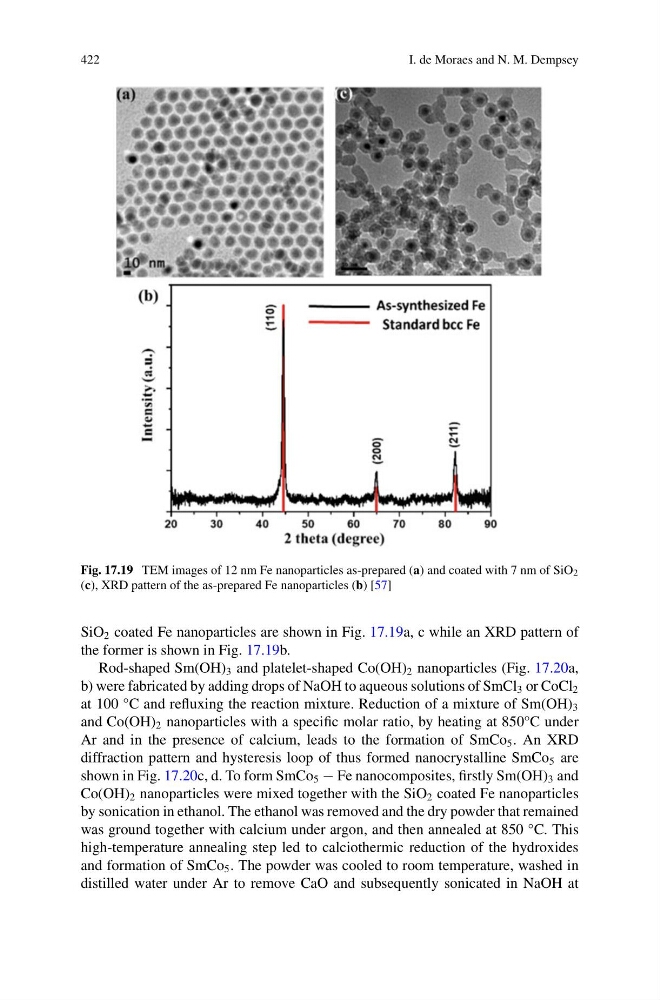
Fig. 17.19 TEM images of 12 nm Fe nanoparticles as-prepared (a) and coated with 7 nm of SiO 2
(c), XRD pattern of the as-prepared Fe nanoparticles (b) [57]
SiO 2 coated Fe nanoparticles are shown in Fig. 17.19a, c while an XRD pattern of
the former is shown in Fig. 17.19b.
Rod-shaped Sm(OH) 3 and platelet-shaped Co(OH) 2 nanoparticles (Fig. 17.20a,
b) were fabricated by adding drops of NaOH to aqueous solutions of SmCl 3 or CoCl 2
at 100 °C and refluxing the reaction mixture. Reduction of a mixture of Sm(OH) 3
and Co(OH) 2 nanoparticles with a specific molar ratio, by heating at 850°C under
Ar and in the presence of calcium, leads to the formation of SmCo 5 . An XRD
diffraction pattern and hysteresis loop of thus formed nanocrystalline SmCo 5 are
shown in Fig. 17.20c, d. To form SmCo 5 − Fe nanocomposites, firstly Sm(OH) 3 and
Co(OH) 2 nanoparticles were mixed together with the SiO 2 coated Fe nanoparticles
by sonication in ethanol. The ethanol was removed and the dry powder that remained
was ground together with calcium under argon, and then annealed at 850 °C. This
high-temperature annealing step led to calciothermic reduction of the hydroxides
and formation of SmCo 5 . The powder was cooled to room temperature, washed in
distilled water under Ar to remove CaO and subsequently sonicated in NaOH at
I. de Moraes and N. M. Dempsey
Fig. 17.19 TEM images of 12 nm Fe nanoparticles as-prepared (a) and coated with 7 nm of SiO 2
(c), XRD pattern of the as-prepared Fe nanoparticles (b) [57]
SiO 2 coated Fe nanoparticles are shown in Fig. 17.19a, c while an XRD pattern of
the former is shown in Fig. 17.19b.
Rod-shaped Sm(OH) 3 and platelet-shaped Co(OH) 2 nanoparticles (Fig. 17.20a,
b) were fabricated by adding drops of NaOH to aqueous solutions of SmCl 3 or CoCl 2
at 100 °C and refluxing the reaction mixture. Reduction of a mixture of Sm(OH) 3
and Co(OH) 2 nanoparticles with a specific molar ratio, by heating at 850°C under
Ar and in the presence of calcium, leads to the formation of SmCo 5 . An XRD
diffraction pattern and hysteresis loop of thus formed nanocrystalline SmCo 5 are
shown in Fig. 17.20c, d. To form SmCo 5 − Fe nanocomposites, firstly Sm(OH) 3 and
Co(OH) 2 nanoparticles were mixed together with the SiO 2 coated Fe nanoparticles
by sonication in ethanol. The ethanol was removed and the dry powder that remained
was ground together with calcium under argon, and then annealed at 850 °C. This
high-temperature annealing step led to calciothermic reduction of the hydroxides
and formation of SmCo 5 . The powder was cooled to room temperature, washed in
distilled water under Ar to remove CaO and subsequently sonicated in NaOH at