

340
M. Avolio et al.
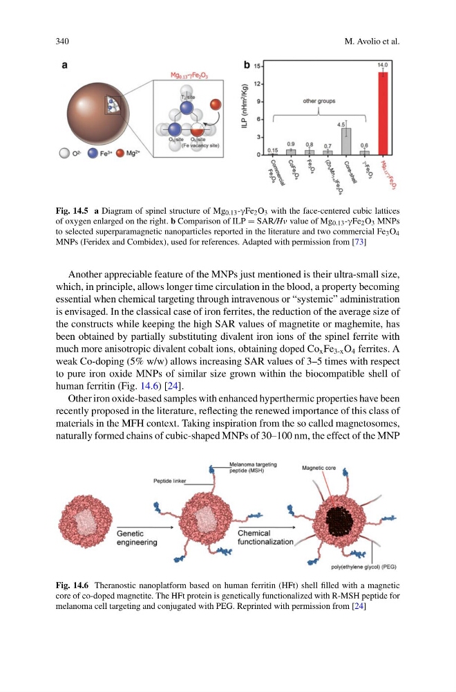
Fig. 14.5 a Diagram of spinel structure of Mg 0.13 -γFe 2 O 3 with the face-centered cubic lattices
of oxygen enlarged on the right. b Comparison of ILP = SAR/Hν value of Mg 0.13 -γFe 2 O 3 MNPs
to selected superparamagnetic nanoparticles reported in the literature and two commercial Fe 3 O 4
MNPs (Feridex and Combidex), used for references. Adapted with permission from [73]
Another appreciable feature of the MNPs just mentioned is their ultra-small size,
which, in principle, allows longer time circulation in the blood, a property becoming
essential when chemical targeting through intravenous or “systemic” administration
is envisaged. In the classical case of iron ferrites, the reduction of the average size of
the constructs while keeping the high SAR values of magnetite or maghemite, has
been obtained by partially substituting divalent iron ions of the spinel ferrite with
much more anisotropic divalent cobalt ions, obtaining doped Co x Fe 3-x O 4 ferrites. A
weak Co-doping (5% w/w) allows increasing SAR values of 3–5 times with respect
to pure iron oxide MNPs of similar size grown within the biocompatible shell of
human ferritin (Fig. 14.6) [24].
Other iron oxide-based samples with enhanced hyperthermic properties have been
recently proposed in the literature, reflecting the renewed importance of this class of
materials in the MFH context. Taking inspiration from the so called magnetosomes,
naturally formed chains of cubic-shaped MNPs of 30–100 nm, the effect of the MNP
Fig. 14.6 Theranostic nanoplatform based on human ferritin (HFt) shell filled with a magnetic
core of co-doped magnetite. The HFt protein is genetically functionalized with R-MSH peptide for
melanoma cell targeting and conjugated with PEG. Reprinted with permission from [24]
M. Avolio et al.
Fig. 14.5 a Diagram of spinel structure of Mg 0.13 -γFe 2 O 3 with the face-centered cubic lattices
of oxygen enlarged on the right. b Comparison of ILP = SAR/Hν value of Mg 0.13 -γFe 2 O 3 MNPs
to selected superparamagnetic nanoparticles reported in the literature and two commercial Fe 3 O 4
MNPs (Feridex and Combidex), used for references. Adapted with permission from [73]
Another appreciable feature of the MNPs just mentioned is their ultra-small size,
which, in principle, allows longer time circulation in the blood, a property becoming
essential when chemical targeting through intravenous or “systemic” administration
is envisaged. In the classical case of iron ferrites, the reduction of the average size of
the constructs while keeping the high SAR values of magnetite or maghemite, has
been obtained by partially substituting divalent iron ions of the spinel ferrite with
much more anisotropic divalent cobalt ions, obtaining doped Co x Fe 3-x O 4 ferrites. A
weak Co-doping (5% w/w) allows increasing SAR values of 3–5 times with respect
to pure iron oxide MNPs of similar size grown within the biocompatible shell of
human ferritin (Fig. 14.6) [24].
Other iron oxide-based samples with enhanced hyperthermic properties have been
recently proposed in the literature, reflecting the renewed importance of this class of
materials in the MFH context. Taking inspiration from the so called magnetosomes,
naturally formed chains of cubic-shaped MNPs of 30–100 nm, the effect of the MNP
Fig. 14.6 Theranostic nanoplatform based on human ferritin (HFt) shell filled with a magnetic
core of co-doped magnetite. The HFt protein is genetically functionalized with R-MSH peptide for
melanoma cell targeting and conjugated with PEG. Reprinted with permission from [24]