

14 Medical Applications of Magnetic Nanoparticles
335
the maximum of SAR grows as the magnetization saturation increases and as the
magneto-crystalline energy decreases, respectively.
The LRT model provides useful indications to foresee the behavior of a magnetic
nanomaterial under the application of an AFM and thus to tune its main features
to optimize the heating process. With this purpose, many other, more sophisticated,
models have been developed in the past years to make the prevision more and more
reliable and thus to address the research on material synthesis toward more effective
devices. Efforts have been made to extend the LRT model by perturbative methods
to include the treatment of larger MNPs, the magnetic behavior of which is at the
borderline between the frequency independent description of the Stoner-Wohlfarth
model and the pure superparamagnetic model referring to the Neél theory [32]. Beside
analytical models to attempt the description of the MNP behavior in this transition
region, computational models, mainly based on Montecarlo simulations, have been
recently developed for accurately evaluating the SAR of a specific nanomaterial as
well as to assess the temperature rise in the surrounding tissues [23, 33].
As established by (14.7), SAR is proportional to the square of the applied field
amplitude, H 0 , and the operating frequency ν. In principle, thus, it might be thought
that the simplest way to increase SAR is to increase the parameters of the external
applied AMF. However, only a narrow window of H 0 and ν are allowed for in vivo
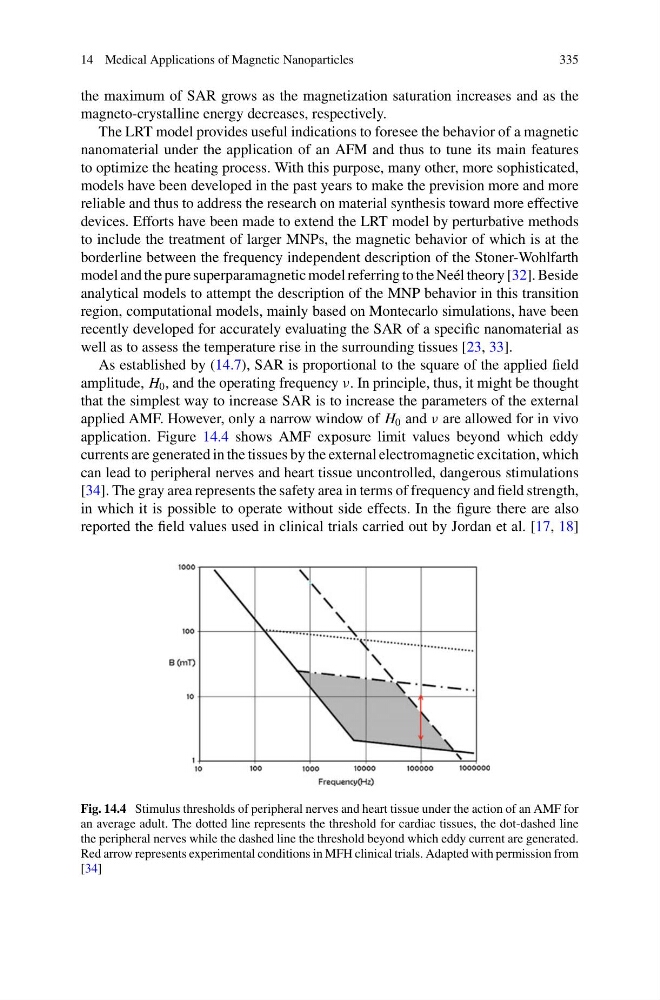
application. Figure 14.4 shows AMF exposure limit values beyond which eddy
currents are generated in the tissues by the external electromagnetic excitation, which
can lead to peripheral nerves and heart tissue uncontrolled, dangerous stimulations
[34]. The gray area represents the safety area in terms of frequency and field strength,
in which it is possible to operate without side effects. In the figure there are also
reported the field values used in clinical trials carried out by Jordan et al. [17, 18]
Fig. 14.4 Stimulus thresholds of peripheral nerves and heart tissue under the action of an AMF for
an average adult. The dotted line represents the threshold for cardiac tissues, the dot-dashed line
the peripheral nerves while the dashed line the threshold beyond which eddy current are generated.
Red arrow represents experimental conditions in MFH clinical trials. Adapted with permission from
[34]
335
the maximum of SAR grows as the magnetization saturation increases and as the
magneto-crystalline energy decreases, respectively.
The LRT model provides useful indications to foresee the behavior of a magnetic
nanomaterial under the application of an AFM and thus to tune its main features
to optimize the heating process. With this purpose, many other, more sophisticated,
models have been developed in the past years to make the prevision more and more
reliable and thus to address the research on material synthesis toward more effective
devices. Efforts have been made to extend the LRT model by perturbative methods
to include the treatment of larger MNPs, the magnetic behavior of which is at the
borderline between the frequency independent description of the Stoner-Wohlfarth
model and the pure superparamagnetic model referring to the Neél theory [32]. Beside
analytical models to attempt the description of the MNP behavior in this transition
region, computational models, mainly based on Montecarlo simulations, have been
recently developed for accurately evaluating the SAR of a specific nanomaterial as
well as to assess the temperature rise in the surrounding tissues [23, 33].
As established by (14.7), SAR is proportional to the square of the applied field
amplitude, H 0 , and the operating frequency ν. In principle, thus, it might be thought
that the simplest way to increase SAR is to increase the parameters of the external
applied AMF. However, only a narrow window of H 0 and ν are allowed for in vivo
application. Figure 14.4 shows AMF exposure limit values beyond which eddy
currents are generated in the tissues by the external electromagnetic excitation, which
can lead to peripheral nerves and heart tissue uncontrolled, dangerous stimulations
[34]. The gray area represents the safety area in terms of frequency and field strength,
in which it is possible to operate without side effects. In the figure there are also
reported the field values used in clinical trials carried out by Jordan et al. [17, 18]
Fig. 14.4 Stimulus thresholds of peripheral nerves and heart tissue under the action of an AMF for
an average adult. The dotted line represents the threshold for cardiac tissues, the dot-dashed line
the peripheral nerves while the dashed line the threshold beyond which eddy current are generated.
Red arrow represents experimental conditions in MFH clinical trials. Adapted with permission from
[34]