

332
M. Avolio et al.
no information about their spatial distribution can be obtained. The main difference
between NMR and MRI techniques is the ability of the latter to give information
on the localization of the nuclei by superimposing to H 0 a combination of gradients
along the three directions, causing a bijective correspondence between magnetic
field value and the nucleus position. These conditions are the basis for the methods
of image reconstruction first described by Lauterbur and Mansfield in 1973, who
were awarded with the Nobel prize in medicine in 2003 [12, 14].
An easy formalism that allows describing the MR image reconstruction is the one
based on the Fourier Transform (FT) operation [29]. First, the signal produced over
the acquisition time by the
1 H spins can be written as
S(t) =
d
3 rρ( r )e
i((t+( r ,t)
(14.2)
where and are the demodulation frequency and the spin phase in a given position
of the space. In presence of field gradients, by introducing three new variables, k x
= γG x t x /2π, k y = γG y t y /2π and k z = γG z t z /2π, it is possible to define the k-space,
which is related to the real space by the FT operation. An equivalent expression for
the signal coming from the nuclei can therefore be written as
S(
k) =
d
3 rρ( r )e
−i2π
k·· r
.
(14.3)
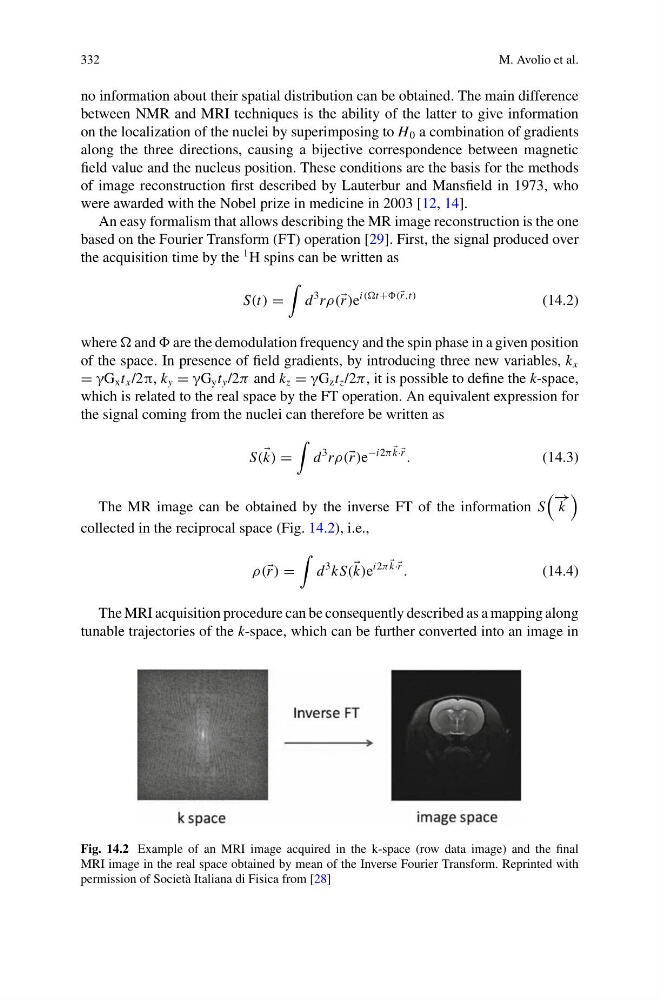
The MR image can be obtained by the inverse FT of the information S
− →
k
collected in the reciprocal space (Fig. 14.2), i.e.,
ρ( r ) =
d
3 kS(
k)e
i2π
k·· r
.
(14.4)
The MRI acquisition procedure can be consequently described as a mapping along
tunable trajectories of the k-space, which can be further converted into an image in
Fig. 14.2 Example of an MRI image acquired in the k-space (row data image) and the final
MRI image in the real space obtained by mean of the Inverse Fourier Transform. Reprinted with
permission of Società Italiana di Fisica from [28]
M. Avolio et al.
no information about their spatial distribution can be obtained. The main difference
between NMR and MRI techniques is the ability of the latter to give information
on the localization of the nuclei by superimposing to H 0 a combination of gradients
along the three directions, causing a bijective correspondence between magnetic
field value and the nucleus position. These conditions are the basis for the methods
of image reconstruction first described by Lauterbur and Mansfield in 1973, who
were awarded with the Nobel prize in medicine in 2003 [12, 14].
An easy formalism that allows describing the MR image reconstruction is the one
based on the Fourier Transform (FT) operation [29]. First, the signal produced over
the acquisition time by the
1 H spins can be written as
S(t) =
d
3 rρ( r )e
i((t+( r ,t)
(14.2)
where and are the demodulation frequency and the spin phase in a given position
of the space. In presence of field gradients, by introducing three new variables, k x
= γG x t x /2π, k y = γG y t y /2π and k z = γG z t z /2π, it is possible to define the k-space,
which is related to the real space by the FT operation. An equivalent expression for
the signal coming from the nuclei can therefore be written as
S(
k) =
d
3 rρ( r )e
−i2π
k·· r
.
(14.3)
The MR image can be obtained by the inverse FT of the information S
− →
k
collected in the reciprocal space (Fig. 14.2), i.e.,
ρ( r ) =
d
3 kS(
k)e
i2π
k·· r
.
(14.4)
The MRI acquisition procedure can be consequently described as a mapping along
tunable trajectories of the k-space, which can be further converted into an image in
Fig. 14.2 Example of an MRI image acquired in the k-space (row data image) and the final
MRI image in the real space obtained by mean of the Inverse Fourier Transform. Reprinted with
permission of Società Italiana di Fisica from [28]