

13 Magnetic Nanoparticles for Life Sciences Applications
311
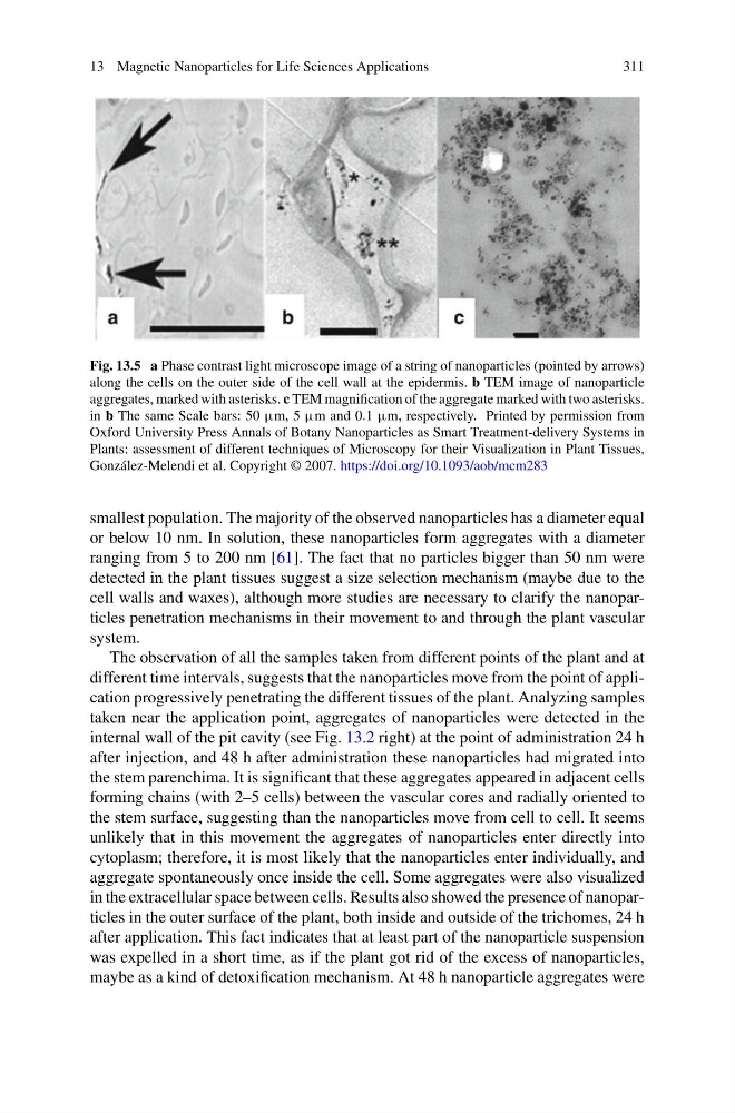
Fig. 13.5 a Phase contrast light microscope image of a string of nanoparticles (pointed by arrows)
along the cells on the outer side of the cell wall at the epidermis. b TEM image of nanoparticle
aggregates, marked with asterisks. c TEM magnification of the aggregate marked with two asterisks.
in b The same Scale bars: 50 μm, 5 μm and 0.1 μm, respectively. Printed by permission from
Oxford University Press Annals of Botany Nanoparticles as Smart Treatment-delivery Systems in
Plants: assessment of different techniques of Microscopy for their Visualization in Plant Tissues,
González-Melendi et al. Copyright © 2007. https://doi.org/10.1093/aob/mcm283
smallest population. The majority of the observed nanoparticles has a diameter equal
or below 10 nm. In solution, these nanoparticles form aggregates with a diameter
ranging from 5 to 200 nm [61]. The fact that no particles bigger than 50 nm were
detected in the plant tissues suggest a size selection mechanism (maybe due to the
cell walls and waxes), although more studies are necessary to clarify the nanoparticles penetration mechanisms in their movement to and through the plant vascular
system.
The observation of all the samples taken from different points of the plant and at
different time intervals, suggests that the nanoparticles move from the point of application progressively penetrating the different tissues of the plant. Analyzing samples
taken near the application point, aggregates of nanoparticles were detected in the
internal wall of the pit cavity (see Fig. 13.2 right) at the point of administration 24 h
after injection, and 48 h after administration these nanoparticles had migrated into
the stem parenchima. It is significant that these aggregates appeared in adjacent cells
forming chains (with 2–5 cells) between the vascular cores and radially oriented to
the stem surface, suggesting than the nanoparticles move from cell to cell. It seems
unlikely that in this movement the aggregates of nanoparticles enter directly into
cytoplasm; therefore, it is most likely that the nanoparticles enter individually, and
aggregate spontaneously once inside the cell. Some aggregates were also visualized
in the extracellular space between cells. Results also showed the presence of nanoparticles in the outer surface of the plant, both inside and outside of the trichomes, 24 h
after application. This fact indicates that at least part of the nanoparticle suspension
was expelled in a short time, as if the plant got rid of the excess of nanoparticles,
maybe as a kind of detoxification mechanism. At 48 h nanoparticle aggregates were
311
Fig. 13.5 a Phase contrast light microscope image of a string of nanoparticles (pointed by arrows)
along the cells on the outer side of the cell wall at the epidermis. b TEM image of nanoparticle
aggregates, marked with asterisks. c TEM magnification of the aggregate marked with two asterisks.
in b The same Scale bars: 50 μm, 5 μm and 0.1 μm, respectively. Printed by permission from
Oxford University Press Annals of Botany Nanoparticles as Smart Treatment-delivery Systems in
Plants: assessment of different techniques of Microscopy for their Visualization in Plant Tissues,
González-Melendi et al. Copyright © 2007. https://doi.org/10.1093/aob/mcm283
smallest population. The majority of the observed nanoparticles has a diameter equal
or below 10 nm. In solution, these nanoparticles form aggregates with a diameter
ranging from 5 to 200 nm [61]. The fact that no particles bigger than 50 nm were
detected in the plant tissues suggest a size selection mechanism (maybe due to the
cell walls and waxes), although more studies are necessary to clarify the nanoparticles penetration mechanisms in their movement to and through the plant vascular
system.
The observation of all the samples taken from different points of the plant and at
different time intervals, suggests that the nanoparticles move from the point of application progressively penetrating the different tissues of the plant. Analyzing samples
taken near the application point, aggregates of nanoparticles were detected in the
internal wall of the pit cavity (see Fig. 13.2 right) at the point of administration 24 h
after injection, and 48 h after administration these nanoparticles had migrated into
the stem parenchima. It is significant that these aggregates appeared in adjacent cells
forming chains (with 2–5 cells) between the vascular cores and radially oriented to
the stem surface, suggesting than the nanoparticles move from cell to cell. It seems
unlikely that in this movement the aggregates of nanoparticles enter directly into
cytoplasm; therefore, it is most likely that the nanoparticles enter individually, and
aggregate spontaneously once inside the cell. Some aggregates were also visualized
in the extracellular space between cells. Results also showed the presence of nanoparticles in the outer surface of the plant, both inside and outside of the trichomes, 24 h
after application. This fact indicates that at least part of the nanoparticle suspension
was expelled in a short time, as if the plant got rid of the excess of nanoparticles,
maybe as a kind of detoxification mechanism. At 48 h nanoparticle aggregates were