

272
P. Torruella et al.
inversion (i.e. cupping artefact) prevent a monotonical behavior of the intensity with
the material composition and, thus, tomography algorithms fail to reconstruct the
SV [37].
One possible solution to overcome this problem is to extract the concentration
of the elements from the intensity edges. Let us consider the case of three elements
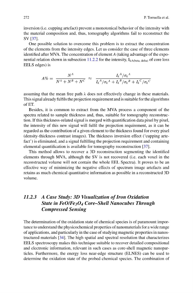
identified after MVA. The concentration of element A (taking advantage of the exponential relation shown in subsection 11.2.2 for the intensity, I kA(beta, delta) of core loss
EELS edges) is
A% =
N
A
N A + N B + N C ≈
I k
A
/σ k
A
I k
A
/σ k
A + I k
B
/σ k
B + I k
C
/σ k
C
assuming that the mean free path λ does not effectively change in these materials.
This signal already fulfils the projection requirement and is suitable for the algorithms
of ET.
Besides, it is common to extract from the MVA process a component of the
spectra related to sample thickness and, thus, suitable for tomography reconstruction. If this thickness-related signal is merged with quantification data pixel by pixel,
the intensity of this new signal will fulfil the projection requirement, as it can be
regarded as the contribution of a given element to the thickness found for every pixel
(density-thickness contrast images). The thickness inversion effect (‘cupping artefact’) is eliminated, and a signal fulfilling the projection requirement and containing
elemental quantification is available for tomography reconstruction [37].
This method allows to recover a 3D reconstruction segmenting the identified
elements through MVA, although the SV is not recovered (i.e. each voxel in the
reconstructed volume will not contain the whole EEL Spectra). It proves to be an
effective way of minimizing the negative effects of spectrum image artefacts and
retains as much chemical quantitative information as possible in a reconstructed 3D
volume.
11.2.3 A Case Study: 3D Visualization of Iron Oxidation
State in FeO/Fe 3 O 4 Core–Shell Nanocubes Through
Compressed Sensing
The determination of the oxidation state of chemical species is of paramount importance to understand the physicochemical properties of nanomaterials for a wide range
of applications, and particularly in the case of studying magnetic properties in nanostructured materials [34]. The high spatial and spectral resolution that characterizes
EELS spectroscopy makes this technique suitable to recover detailed compositional
and electronic information, relevant in such cases as core-shell magnetic nanoparticles. Furthermore, the energy loss near-edge structure (ELNES) can be used to
determine the oxidation state of the probed chemical species. The combination of
P. Torruella et al.
inversion (i.e. cupping artefact) prevent a monotonical behavior of the intensity with
the material composition and, thus, tomography algorithms fail to reconstruct the
SV [37].
One possible solution to overcome this problem is to extract the concentration
of the elements from the intensity edges. Let us consider the case of three elements
identified after MVA. The concentration of element A (taking advantage of the exponential relation shown in subsection 11.2.2 for the intensity, I kA(beta, delta) of core loss
EELS edges) is
A% =
N
A
N A + N B + N C ≈
I k
A
/σ k
A
I k
A
/σ k
A + I k
B
/σ k
B + I k
C
/σ k
C
assuming that the mean free path λ does not effectively change in these materials.
This signal already fulfils the projection requirement and is suitable for the algorithms
of ET.
Besides, it is common to extract from the MVA process a component of the
spectra related to sample thickness and, thus, suitable for tomography reconstruction. If this thickness-related signal is merged with quantification data pixel by pixel,
the intensity of this new signal will fulfil the projection requirement, as it can be
regarded as the contribution of a given element to the thickness found for every pixel
(density-thickness contrast images). The thickness inversion effect (‘cupping artefact’) is eliminated, and a signal fulfilling the projection requirement and containing
elemental quantification is available for tomography reconstruction [37].
This method allows to recover a 3D reconstruction segmenting the identified
elements through MVA, although the SV is not recovered (i.e. each voxel in the
reconstructed volume will not contain the whole EEL Spectra). It proves to be an
effective way of minimizing the negative effects of spectrum image artefacts and
retains as much chemical quantitative information as possible in a reconstructed 3D
volume.
11.2.3 A Case Study: 3D Visualization of Iron Oxidation
State in FeO/Fe 3 O 4 Core–Shell Nanocubes Through
Compressed Sensing
The determination of the oxidation state of chemical species is of paramount importance to understand the physicochemical properties of nanomaterials for a wide range
of applications, and particularly in the case of studying magnetic properties in nanostructured materials [34]. The high spatial and spectral resolution that characterizes
EELS spectroscopy makes this technique suitable to recover detailed compositional
and electronic information, relevant in such cases as core-shell magnetic nanoparticles. Furthermore, the energy loss near-edge structure (ELNES) can be used to
determine the oxidation state of the probed chemical species. The combination of