

242
C. Binns et al.
in K-edge absorption for left and right circularly polarised photons [2]. As shown
by Erskine and Stern, however, in the case of transition metals, L 2,3 -edge absorption
in the soft X-ray band is potentially a more powerful source of information though
technically more difficult. An important breakthrough starting in the early 1980s was
the emergence of dedicated synchrotron radiation sources that could produce high
intensities of circularly polarised soft X-rays.
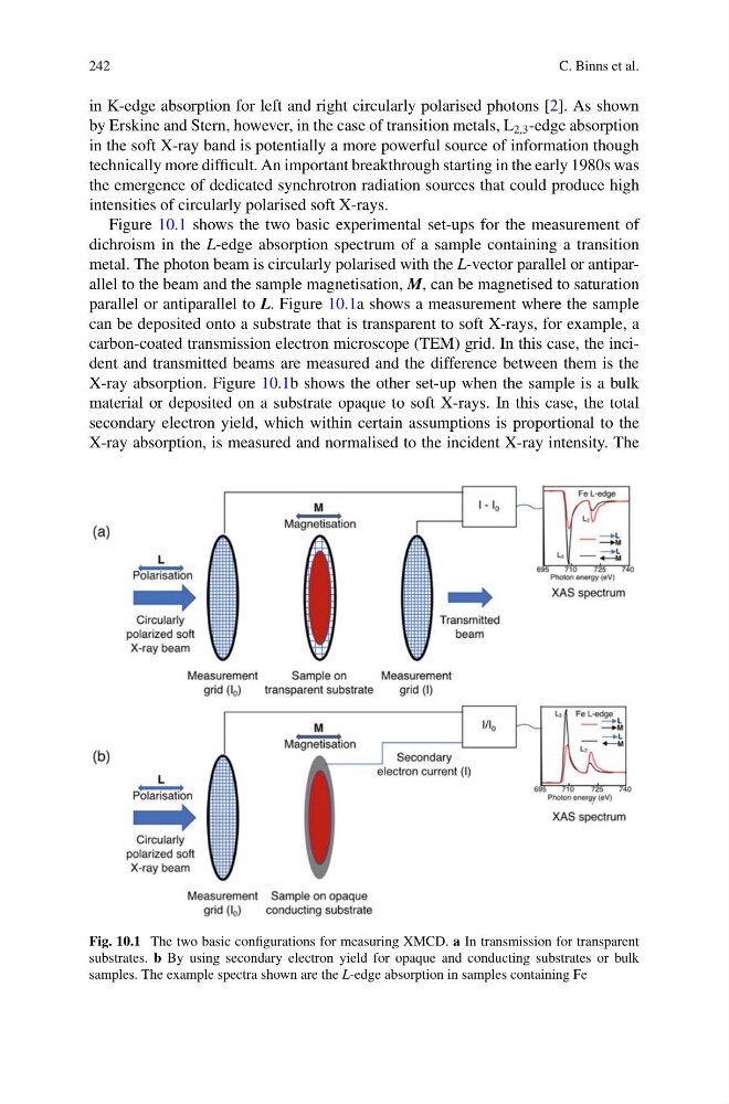
Figure 10.1 shows the two basic experimental set-ups for the measurement of
dichroism in the L-edge absorption spectrum of a sample containing a transition
metal. The photon beam is circularly polarised with the L-vector parallel or antiparallel to the beam and the sample magnetisation, M, can be magnetised to saturation
parallel or antiparallel to L. Figure 10.1a shows a measurement where the sample
can be deposited onto a substrate that is transparent to soft X-rays, for example, a
carbon-coated transmission electron microscope (TEM) grid. In this case, the incident and transmitted beams are measured and the difference between them is the
X-ray absorption. Figure 10.1b shows the other set-up when the sample is a bulk
material or deposited on a substrate opaque to soft X-rays. In this case, the total
secondary electron yield, which within certain assumptions is proportional to the
X-ray absorption, is measured and normalised to the incident X-ray intensity. The
Fig. 10.1 The two basic configurations for measuring XMCD. a In transmission for transparent
substrates. b By using secondary electron yield for opaque and conducting substrates or bulk
samples. The example spectra shown are the L-edge absorption in samples containing Fe
C. Binns et al.
in K-edge absorption for left and right circularly polarised photons [2]. As shown
by Erskine and Stern, however, in the case of transition metals, L 2,3 -edge absorption
in the soft X-ray band is potentially a more powerful source of information though
technically more difficult. An important breakthrough starting in the early 1980s was
the emergence of dedicated synchrotron radiation sources that could produce high
intensities of circularly polarised soft X-rays.
Figure 10.1 shows the two basic experimental set-ups for the measurement of
dichroism in the L-edge absorption spectrum of a sample containing a transition
metal. The photon beam is circularly polarised with the L-vector parallel or antiparallel to the beam and the sample magnetisation, M, can be magnetised to saturation
parallel or antiparallel to L. Figure 10.1a shows a measurement where the sample
can be deposited onto a substrate that is transparent to soft X-rays, for example, a
carbon-coated transmission electron microscope (TEM) grid. In this case, the incident and transmitted beams are measured and the difference between them is the
X-ray absorption. Figure 10.1b shows the other set-up when the sample is a bulk
material or deposited on a substrate opaque to soft X-rays. In this case, the total
secondary electron yield, which within certain assumptions is proportional to the
X-ray absorption, is measured and normalised to the incident X-ray intensity. The
Fig. 10.1 The two basic configurations for measuring XMCD. a In transmission for transparent
substrates. b By using secondary electron yield for opaque and conducting substrates or bulk
samples. The example spectra shown are the L-edge absorption in samples containing Fe