

7 Nature Driven Magnetic Nanoarchitectures
165
20 60 100 140 180 220 260 300 340 380
0.0
0.5
1.0
1.5
2.0
2.5
mass of Fe (fg/cell)
time (min)
ferrihydrite
magnetite
0
0.5
1
1.5
7100
7120
7140
7160
7180
7200
20 min
60 min
100 min
140 min
180 min
240 min
360 min
absorption
Energy (eV)
7110
7120
0
0.2
0.4
(a)
(b)
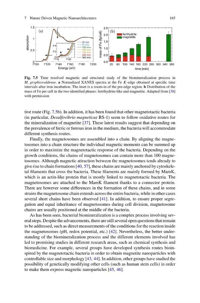
Fig. 7.5 Time resolved magnetic and structural study of the biomineralization process in
M. gryphiswaldense. a Normalized XANES spectra at the Fe K -edge obtained at specific time
intervals after iron incubation. The inset is a zoom-in of the pre-edge region. b Distribution of the
mass of Fe per cell in the two identified phases: ferrihydrite-like and magnetite. Adapted from [38]
with permission
tive route (Fig. 7.5b). In addition, it has been found that other magnetotactic bacteria
(in particular, Desulfovibrio magneticus RS-1) seem to follow oxidative routes for
the mineralization of magnetite [37]. These latest results suggest that depending on
the prevalence of ferric or ferrous iron in the medium, the bacteria will accommodate
different synthesis routes.
Finally, the magnetosomes are assembled into a chain. By aligning the magnetosomes into a chain structure the individual magnetic moments can be summed up
in order to maximize the magnetotactic response of the bacteria. Depending on the
growth conditions, the chains of magnetosomes can contain more than 100 magnetosomes. Although magnetic attraction between the magnetosomes tends already to
give rise to chain formations [40, 57], these chains are mainly anchored by cytoskeletal filaments that cross the bacteria. These filaments are mainly formed by MamK,
which is an actin-like protein that is mostly linked to magnetotactic bacteria. The
magnetosomes are attached to the MamK filament thanks to a connector protein.
There are however some differences in the formation of these chains, and in some
strains the magnetosome chain extends across the entire bacteria, while in other cases
several short chains have been observed [41]. In addition, to ensure proper segregation and equal inheritance of magnetosomes during cell division, magnetosome
chains are usually positioned at the middle of the bacteria.
As has been seen, bacterial biomineralization is a complex process involving several steps. Despite the advancements, there are still several open questions that remain
to be addressed, such as direct measurements of the conditions for the reaction inside
the magnetosomes (pH, redox potential, etc.) [42]. Nevertheless, the better understanding of the biomineralization process and the different elements involved has
led to promising studies in different research areas, such as chemical synthesis and
biomedicine. For example, several groups have developed synthesis routes bioinspired by the magnetotactic bacteria in order to obtain magnetite nanoparticles with
controllable size and morphology [43, 44]. In addition, other groups have studied the
possibility of genetically modifying other cells (such as human stem cells) in order
to make them express magnetic nanoparticles [45, 46].
165
20 60 100 140 180 220 260 300 340 380
0.0
0.5
1.0
1.5
2.0
2.5
mass of Fe (fg/cell)
time (min)
ferrihydrite
magnetite
0
0.5
1
1.5
7100
7120
7140
7160
7180
7200
20 min
60 min
100 min
140 min
180 min
240 min
360 min
absorption
Energy (eV)
7110
7120
0
0.2
0.4
(a)
(b)
Fig. 7.5 Time resolved magnetic and structural study of the biomineralization process in
M. gryphiswaldense. a Normalized XANES spectra at the Fe K -edge obtained at specific time
intervals after iron incubation. The inset is a zoom-in of the pre-edge region. b Distribution of the
mass of Fe per cell in the two identified phases: ferrihydrite-like and magnetite. Adapted from [38]
with permission
tive route (Fig. 7.5b). In addition, it has been found that other magnetotactic bacteria
(in particular, Desulfovibrio magneticus RS-1) seem to follow oxidative routes for
the mineralization of magnetite [37]. These latest results suggest that depending on
the prevalence of ferric or ferrous iron in the medium, the bacteria will accommodate
different synthesis routes.
Finally, the magnetosomes are assembled into a chain. By aligning the magnetosomes into a chain structure the individual magnetic moments can be summed up
in order to maximize the magnetotactic response of the bacteria. Depending on the
growth conditions, the chains of magnetosomes can contain more than 100 magnetosomes. Although magnetic attraction between the magnetosomes tends already to
give rise to chain formations [40, 57], these chains are mainly anchored by cytoskeletal filaments that cross the bacteria. These filaments are mainly formed by MamK,
which is an actin-like protein that is mostly linked to magnetotactic bacteria. The
magnetosomes are attached to the MamK filament thanks to a connector protein.
There are however some differences in the formation of these chains, and in some
strains the magnetosome chain extends across the entire bacteria, while in other cases
several short chains have been observed [41]. In addition, to ensure proper segregation and equal inheritance of magnetosomes during cell division, magnetosome
chains are usually positioned at the middle of the bacteria.
As has been seen, bacterial biomineralization is a complex process involving several steps. Despite the advancements, there are still several open questions that remain
to be addressed, such as direct measurements of the conditions for the reaction inside
the magnetosomes (pH, redox potential, etc.) [42]. Nevertheless, the better understanding of the biomineralization process and the different elements involved has
led to promising studies in different research areas, such as chemical synthesis and
biomedicine. For example, several groups have developed synthesis routes bioinspired by the magnetotactic bacteria in order to obtain magnetite nanoparticles with
controllable size and morphology [43, 44]. In addition, other groups have studied the
possibility of genetically modifying other cells (such as human stem cells) in order
to make them express magnetic nanoparticles [45, 46].