

164
M. L. Fdez-Gubieda et al.
MamJ
Mms
Outer Membrane
Inner Membrane
Membrane
InvaginaƟon
Iron uptake
Iron
transportaƟon
MagneƟte
nucleaƟon
MagneƟte
growth
Extracellular
Fe 3+ or Fe 2+
Bfr-like
MagneƟte
(Fe 3+ )
(Fe 2+ ,Fe 3+ )
Ferrihydrite-like
structure (Fe 3+ )
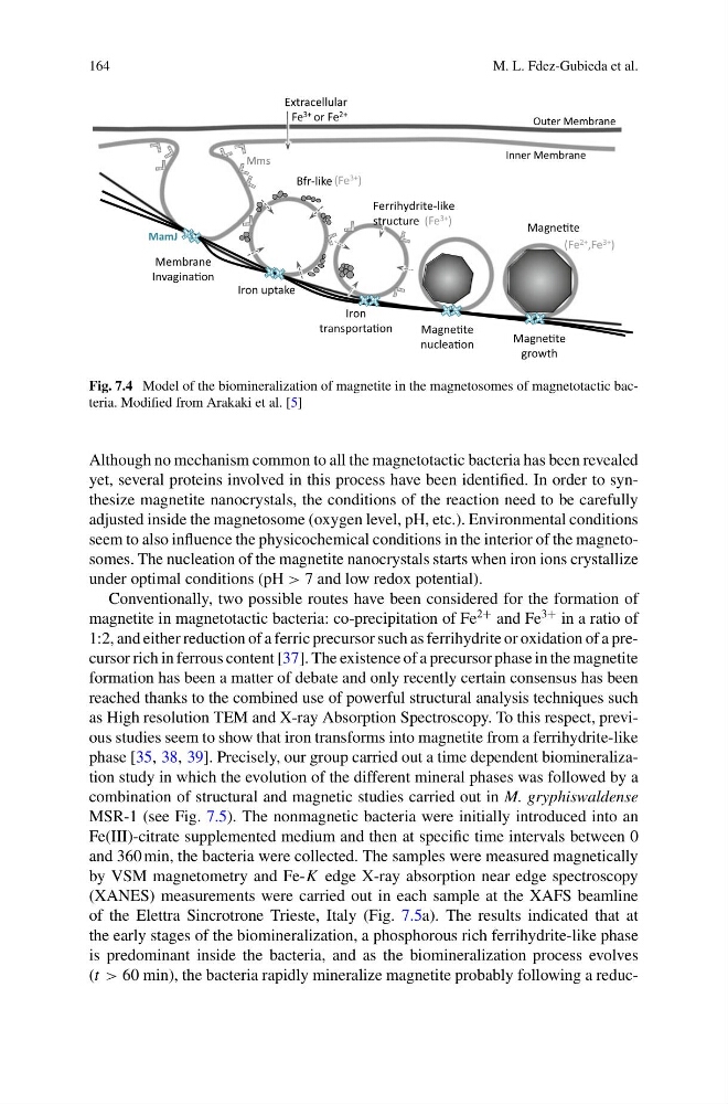
Fig. 7.4 Model of the biomineralization of magnetite in the magnetosomes of magnetotactic bacteria. Modified from Arakaki et al. [5]
Although no mechanism common to all the magnetotactic bacteria has been revealed
yet, several proteins involved in this process have been identified. In order to synthesize magnetite nanocrystals, the conditions of the reaction need to be carefully
adjusted inside the magnetosome (oxygen level, pH, etc.). Environmental conditions
seem to also influence the physicochemical conditions in the interior of the magnetosomes. The nucleation of the magnetite nanocrystals starts when iron ions crystallize
under optimal conditions (pH > 7 and low redox potential).
Conventionally, two possible routes have been considered for the formation of
magnetite in magnetotactic bacteria: co-precipitation of Fe
2+ and Fe
3+ in a ratio of
1:2, and either reduction of a ferric precursor such as ferrihydrite or oxidation of a precursor rich in ferrous content [37]. The existence of a precursor phase in the magnetite
formation has been a matter of debate and only recently certain consensus has been
reached thanks to the combined use of powerful structural analysis techniques such
as High resolution TEM and X-ray Absorption Spectroscopy. To this respect, previous studies seem to show that iron transforms into magnetite from a ferrihydrite-like
phase [35, 38, 39]. Precisely, our group carried out a time dependent biomineralization study in which the evolution of the different mineral phases was followed by a
combination of structural and magnetic studies carried out in M. gryphiswaldense
MSR-1 (see Fig. 7.5). The nonmagnetic bacteria were initially introduced into an
Fe(III)-citrate supplemented medium and then at specific time intervals between 0
and 360 min, the bacteria were collected. The samples were measured magnetically
by VSM magnetometry and Fe-K edge X-ray absorption near edge spectroscopy
(XANES) measurements were carried out in each sample at the XAFS beamline
of the Elettra Sincrotrone Trieste, Italy (Fig. 7.5a). The results indicated that at
the early stages of the biomineralization, a phosphorous rich ferrihydrite-like phase
is predominant inside the bacteria, and as the biomineralization process evolves
(t > 60 min), the bacteria rapidly mineralize magnetite probably following a reduc-
M. L. Fdez-Gubieda et al.
MamJ
Mms
Outer Membrane
Inner Membrane
Membrane
InvaginaƟon
Iron uptake
Iron
transportaƟon
MagneƟte
nucleaƟon
MagneƟte
growth
Extracellular
Fe 3+ or Fe 2+
Bfr-like
MagneƟte
(Fe 3+ )
(Fe 2+ ,Fe 3+ )
Ferrihydrite-like
structure (Fe 3+ )
Fig. 7.4 Model of the biomineralization of magnetite in the magnetosomes of magnetotactic bacteria. Modified from Arakaki et al. [5]
Although no mechanism common to all the magnetotactic bacteria has been revealed
yet, several proteins involved in this process have been identified. In order to synthesize magnetite nanocrystals, the conditions of the reaction need to be carefully
adjusted inside the magnetosome (oxygen level, pH, etc.). Environmental conditions
seem to also influence the physicochemical conditions in the interior of the magnetosomes. The nucleation of the magnetite nanocrystals starts when iron ions crystallize
under optimal conditions (pH > 7 and low redox potential).
Conventionally, two possible routes have been considered for the formation of
magnetite in magnetotactic bacteria: co-precipitation of Fe
2+ and Fe
3+ in a ratio of
1:2, and either reduction of a ferric precursor such as ferrihydrite or oxidation of a precursor rich in ferrous content [37]. The existence of a precursor phase in the magnetite
formation has been a matter of debate and only recently certain consensus has been
reached thanks to the combined use of powerful structural analysis techniques such
as High resolution TEM and X-ray Absorption Spectroscopy. To this respect, previous studies seem to show that iron transforms into magnetite from a ferrihydrite-like
phase [35, 38, 39]. Precisely, our group carried out a time dependent biomineralization study in which the evolution of the different mineral phases was followed by a
combination of structural and magnetic studies carried out in M. gryphiswaldense
MSR-1 (see Fig. 7.5). The nonmagnetic bacteria were initially introduced into an
Fe(III)-citrate supplemented medium and then at specific time intervals between 0
and 360 min, the bacteria were collected. The samples were measured magnetically
by VSM magnetometry and Fe-K edge X-ray absorption near edge spectroscopy
(XANES) measurements were carried out in each sample at the XAFS beamline
of the Elettra Sincrotrone Trieste, Italy (Fig. 7.5a). The results indicated that at
the early stages of the biomineralization, a phosphorous rich ferrihydrite-like phase
is predominant inside the bacteria, and as the biomineralization process evolves
(t > 60 min), the bacteria rapidly mineralize magnetite probably following a reduc-